
Rabbit Anti-phospho-Smad2/Smad3 (Thr8)antibody
Smad2 + Smad3 (phospho T8); p-Smad2 + Smad3 (phospho T8); P-Smad2 /3 (phospho T8); hMAD 2; hMAD 3; hSMAD2; hSMAD3; Mad related protein 2; MADH2; MADH3; MADR2; Mothers against DPP homolog 2; Mothers against DPP homolog 3; Sma and Mad related protein 2; SMA
View History [Clear]
Details
Product Name phospho-Smad2/Smad3 (Thr8) Chinese Name 磷酸化细胞Signal transduction分子SMAD2/SMAD3抗体 Alias Smad2 + Smad3 (phospho T8); p-Smad2 + Smad3 (phospho T8); P-Smad2 /3 (phospho T8); hMAD 2; hMAD 3; hSMAD2; hSMAD3; Mad related protein 2; MADH2; MADH3; MADR2; Mothers against DPP homolog 2; Mothers against DPP homolog 3; Sma and Mad related protein 2; SMA and MAD related protein 3; SMAD 2; SMAD 3; SMAD family member 2; SMAD family member 3. literatures Product Type Phosphorylated anti Research Area Tumour Cell biology immunology Signal transduction Apoptosis transcriptional regulatory factor Epigenetics Immunogen Species Rabbit Clonality Polyclonal React Species Human, Mouse, (predicted: Rat, Chicken, Dog, Pig, Cow, Horse, ) Applications WB=1:500-2000 ELISA=1:5000-10000 IHC-P=1:100-500 IHC-F=1:100-500 Flow-Cyt=1μg/Test ICC=1:100 IF=1:100-500 (Paraffin sections need antigen repair)
not yet tested in other applications.
optimal dilutions/concentrations should be determined by the end user.Theoretical molecular weight 52kDa Cellular localization The nucleus cytoplasmic Form Liquid Concentration 1mg/ml immunogen KLH conjugated synthesised phosphopeptide derived from human Smad2/Smad3 around the phosphorylation site of Thr8: PF(p-T)PP Lsotype IgG Purification affinity purified by Protein A Buffer Solution 0.01M TBS(pH7.4) with 1% BSA, 0.03% Proclin300 and 50% Glycerol. Storage Shipped at 4℃. Store at -20 °C for one year. Avoid repeated freeze/thaw cycles. Attention This product as supplied is intended for research use only, not for use in human, therapeutic or diagnostic applications. PubMed PubMed Product Detail The protein encoded by this gene belongs to the SMAD, a family of proteins similar to the gene products of the Drosophila gene 'mothers against decapentaplegic' (Mad) and the C. elegans gene Sma. SMAD proteins are signal transducers and transcriptional modulators that mediate multiple signaling pathways. This protein mediates the signal of the transforming growth factor (TGF)-beta, and thus regulates multiple cellular processes, such as cell proliferation, apoptosis, and differentiation. This protein is recruited to the TGF-beta receptors through its interaction with the SMAD anchor for receptor activation (SARA) protein. In response to TGF-beta signal, this protein is phosphorylated by the TGF-beta receptors. The phosphorylation induces the dissociation of this protein with SARA and the association with the family member SMAD4. The association with SMAD4 is important for the translocation of this protein into the nucleus, where it binds to target promoters and forms a transcription repressor complex with other cofactors. This protein can also be phosphorylated by activin type 1 receptor kinase, and mediates the signal from the activin. Alternatively spliced transcript variants have been observed for this gene. [provided by RefSeq, May 2012]
Function:
SMAD is a family of proteins similar to the gene products of the Drosophila gene 'mothers against decapentaplegic' (Mad) and the C. elegans gene Sma. SMAD proteins are signal transducers and transcriptional modulators that mediate multiple signaling pathways. They mediate the signal of the transforming growth factor (TGF)-beta, and thus regulate multiple cellular processes, such as cell proliferation, apoptosis, and differentiation.
Subcellular Location:
Cytoplasm. Nucleus. Note: Cytoplasmic in the absence of ligand. Migrates to the nucleus when complexed with SMAD4.
SWISS:
P84022
Gene ID:
4087
Database links:Entrez Gene: 4087 Human
Entrez Gene: 4088 Human
Entrez Gene: 17126 Mouse
Entrez Gene: 17127 Mouse
Omim: 601366 Human
Omim: 603109 Human
SwissProt: P84022 Human
SwissProt: Q15796 Human
SwissProt: Q62432 Mouse
SwissProt: Q8BUN5 Mouse
Unigene: 12253 Human
Unigene: 714621 Human
Unigene: 10636 Rat
Product Picture 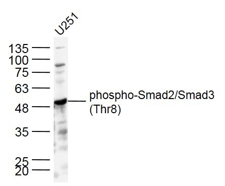 Sample:
U251 cell(human) Lysate at 30 ug
Primary: Anti- p-Smad2/Smad3 (Thr8) (SL8853R) at 1/500 dilution
Secondary: IRDye800CW Goat Anti-Rabbit IgG at 1/20000 dilution
Predicted band size: 52kD
Observed band size: 52 kD
 Sample:
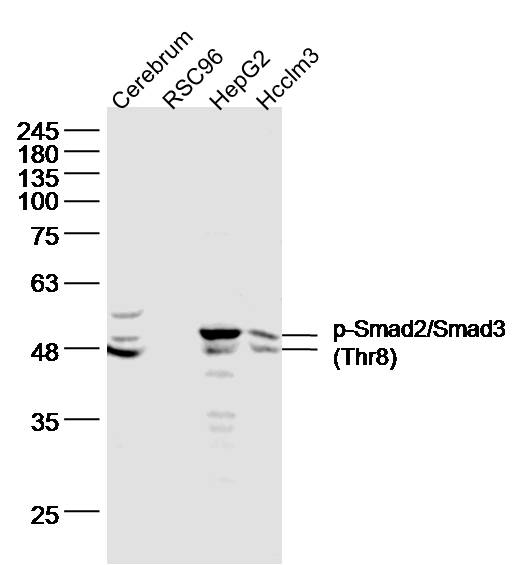
cerebrum(mouse) Lysate at 40 ug
RSC96 cell(rat) Lysate at 30 ug
hepG2 cell(human) Lysate at 30 ug
Hcclm3 cell(human) Lysate at 30 ug
Primary: Anti- p-Smad2/Smad3 (Thr8) (SL8853R) at 1/500 dilution
Secondary: IRDye800CW Goat Anti-Rabbit IgG at 1/20000 dilution
Predicted band size: 52kD
Observed band size: 48,52 kD
 Sample:
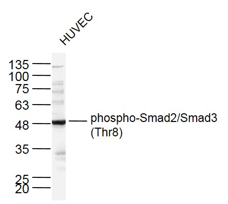
HUVEC cell(human) Lysate at 30 ug
Primary: Anti- p-Smad2/Smad3 (Thr8) (SL8853R) at 1/500 dilution
Secondary: IRDye800CW Goat Anti-Rabbit IgG at 1/20000 dilution
Predicted band size: 52kD
Observed band size: 52 kD
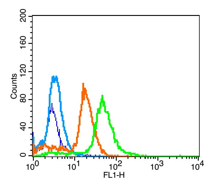 blank: A549 cells (blue line)
isotype control: rabbit IgG (orange line)
second antibody: goat anti-rabbit IgG (white blue line)
primary antibody: rabbit Anti-phospho-Smad2/Smad3 (Thr8) (green line); contration: 3μg /10^6 cells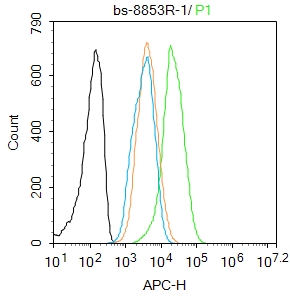 Blank control: Hela.
Primary Antibody (green line): Rabbit Anti-phospho-Smad2/Smad3 (Thr8) antibody (SL8853R)
Dilution: 1μg /10^6 cells;
Isotype Control Antibody (orange line): Rabbit IgG .
Secondary Antibody : Goat anti-rabbit IgG-AF647
Dilution: 1μg /test.
Protocol
The cells were fixed with 4% PFA (10min at room temperature)and then permeabilized with 90% ice-cold methanol for 20 min at -20℃. The cells were then incubated in 5%BSA to block non-specific protein-protein interactions for 30 min at room temperature .Cells stained with Primary Antibody for 30 min at room temperature. The secondary antibody used for 40 min at room temperature. Acquisition of 20,000 events was performed.
Cartpieces
Totalgoods,subtotals:¥Checkout
References (0)
No References
Bought notes(bought amounts latest0)
No one bought this product
User Comment(Total0User Comment Num)
- No comment


 +86 571 56623320
 +86 18668110335

