
Rabbit Anti-CLEC 4E antibody
C type lectin domain family 4 member E; C type lectin superfamily member 9; C-type (calcium dependent carbohydrate recognition domain) lectin superfamily member 9; C-type lectin domain family 4 member E; C-type lectin superfamily member 9; CLC4E_HUMAN; CL
View History [Clear]
Details
Product Name CLEC 4E Chinese Name C型凝集素4家族E抗体 Alias C type lectin domain family 4 member E; C type lectin superfamily member 9; C-type (calcium dependent carbohydrate recognition domain) lectin superfamily member 9; C-type lectin domain family 4 member E; C-type lectin superfamily member 9; CLC4E_HUMAN; CLEC 4E; CLEC4E Clec4e; CLECSF9; Macrophage inducible C type lectin; Macrophage-inducible C-type lectin; MINCLE. literatures Research Area Cell biology Signal transduction Apoptosis Immunogen Species Rabbit Clonality Polyclonal React Species Human, (predicted: Mouse, Rat, Cow, Horse, ) Applications ELISA=1:5000-10000 IHC-P=1:100-500 IHC-F=1:100-500 Flow-Cyt=2μg/Test IF=1:50-200 (Paraffin sections need antigen repair)
not yet tested in other applications.
optimal dilutions/concentrations should be determined by the end user.Theoretical molecular weight 25kDa Cellular localization The cell membrane Form Liquid Concentration 1mg/ml immunogen KLH conjugated synthetic peptide derived from human CLECSF9: 51-150/219 <Extracellular> Lsotype IgG Purification affinity purified by Protein A Buffer Solution 0.01M TBS(pH7.4) with 1% BSA, 0.03% Proclin300 and 50% Glycerol. Storage Shipped at 4℃. Store at -20 °C for one year. Avoid repeated freeze/thaw cycles. Attention This product as supplied is intended for research use only, not for use in human, therapeutic or diagnostic applications. PubMed PubMed Product Detail C-type lectin that functions as cell-surface receptor for a wide variety of ligands such as damaged cells, fungi and mycobacteria. Plays a role in the recognition of pathogenic fungi, such as Candida albicans. The detection of mycobacteria is via trehalose 6,6'-dimycolate (TDM), a cell wall glycolipid. Specifically recognizes alpha-mannose residues on pathogenic fungi of the genus Malassezia. Recognizes also SAP130, a nuclear protein, that is released by dead or dying cells. Transduces signals through an ITAM-containing adapter protein, Fc receptor gamma chain /FCER1G. Induces secretion of inflammatory cytokines through a pathway that depends on SYK, CARD9 and NF-kappa-B.
Function:
C-type lectin that functions as cell-surface receptor for a wide variety of ligands such as damaged cells, fungi and mycobacteria. Plays a role in the recognition of pathogenic fungi, such as Candida albicans. The detection of mycobacteria is via trehalose 6,6'-dimycolate (TDM), a cell wall glycolipid. Specifically recognizes alpha-mannose residues on pathogenic fungi of the genus Malassezia. Recognizes also SAP130, a nuclear protein, that is released by dead or dying cells. Transduces signals through an ITAM-containing adapter protein, Fc receptor gamma chain /FCER1G. Induces secretion of inflammatory cytokines through a pathway that depends on SYK, CARD9 and NF-kappa-B.
Subunit:
Monomer. Homodimer.
Subcellular Location:
Membrane.
Similarity:
Contains 1 C-type lectin domain.
SWISS:
Q9ULY5
Gene ID:
26253
Database links:Entrez Gene: 26253 Human
Omim: 609962 Human
SwissProt: Q9ULY5 Human
Unigene: 236516 Human
Product Picture 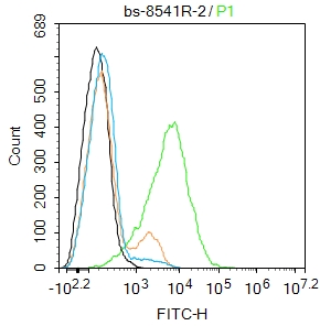 Blank control:THP-1.
Primary Antibody (green line): Rabbit Anti-CLEC 4E antibody (SL8541R)
Dilution: 2μg /10^6 cells;
Isotype Control Antibody (orange line): Rabbit IgG .
Secondary Antibody : Goat anti-rabbit IgG-FITC
Dilution: 0.5μg /test.
Protocol
The cells were incubated in 5%BSA to block non-specific protein-protein interactions for 30 min at room temperature .Cells stained with Primary Antibody for 30 min at room temperature. The secondary antibody used for 40 min at room temperature. Acquisition of 20,000 events was performed.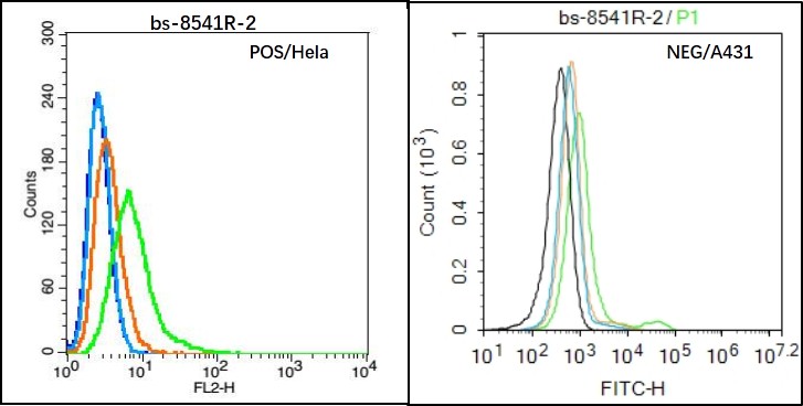 Black line : Positive blank control (Hela); Negative blank control (A431)
Green line : Primary Antibody (Rabbit Anti-CLEC 4E antibody (SL8541R) )
Orange line:Isotype Control Antibody (Rabbit IgG) .
Blue line : Secondary Antibody (Goat anti-rabbit IgG-PE)/(Goat anti-rabbit IgG-AF488)
Hela(Positive)and A431(Negative control)cells (black) were incubated in 5% BSA blocking buffer for 30 min at room temperature. Cells were then stained with CLEC 4E Antibody(SL8541R)at 1:50 dilution in blocking buffer and incubated for 30 min at room temperature, washed twice with 2% BSA in PBS, followed by secondary antibody(blue) incubation for 40 min at room temperature. Acquisitions of 20,000 events were performed. Cells stained with primary antibody (green), and isotype control (orange).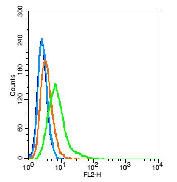 Blank control: Hela(blue), the cells were fixed with 2% paraformaldehyde (10 min) and then permeabilized with ice-cold 90% methanol for 30 min on ice.
Isotype Control Antibody: Rabbit IgG(orange) ; Secondary Antibody: Goat anti-rabbit IgG-PE(white blue), Dilution: 1:200 in 1 X PBS containing 0.5% BSA ; Primary Antibody Dilution: 5μg in 100 μL1X PBS containing 0.5% BSA(green).
Cartpieces
Totalgoods,subtotals:¥Checkout
References (0)
No References
Bought notes(bought amounts latest0)
No one bought this product
User Comment(Total0User Comment Num)
- No comment


 +86 571 56623320
 +86 18668110335

