
Rabbit Anti-caspase-9 p10 antibody
APAF 3; APAF3; Apoptosis related cysteine peptidase; Apoptotic protease activating factor 3; Apoptotic protease MCH 6; Apoptotic protease MCH6 antibodyCASP 9; CASP9; Caspase 9; Caspase 9 apoptosis related cysteine protease; Caspase 9 precursor; Caspase 9
View History [Clear]
Details
Product Name caspase-9 p10 Chinese Name 半胱胺酸蛋白酶蛋白9-p10抗体 Alias APAF 3; APAF3; Apoptosis related cysteine peptidase; Apoptotic protease activating factor 3; Apoptotic protease MCH 6; Apoptotic protease MCH6 antibody CASP 9; CASP9; Caspase 9; Caspase 9 apoptosis related cysteine protease; Caspase 9 precursor; Caspase 9 subunit p10; Caspase 9c; Caspase9; Caspase9 subunit p10; ICE LAP6; ICE like apoptotic protease 6; MCH 6; MCH6; OTTHUMP00000044594; CASP9_HUMAN. literatures Research Area Cell biology immunology Neurobiology Apoptosis Immunogen Species Rabbit Clonality Polyclonal React Species Human, Mouse, Rat, (predicted: Chicken, Dog, Pig, Cow, Horse, Rabbit, ) Applications WB=1:500-2000 ELISA=1:5000-10000 IHC-P=1:100-500 IHC-F=1:100-500 Flow-Cyt=1μg /Test IF=1:50-200 (Paraffin sections need antigen repair)
not yet tested in other applications.
optimal dilutions/concentrations should be determined by the end user.Theoretical molecular weight 10/50 kDa Cellular localization Secretory protein Form Liquid Concentration 1mg/ml immunogen KLH conjugated synthetic peptide derived from human caspase-9 subunit p10: 351-416/416 Lsotype IgG Purification affinity purified by Protein A Buffer Solution 0.01M TBS(pH7.4) with 1% BSA, 0.03% Proclin300 and 50% Glycerol. Storage Shipped at 4℃. Store at -20 °C for one year. Avoid repeated freeze/thaw cycles. Attention This product as supplied is intended for research use only, not for use in human, therapeutic or diagnostic applications. PubMed PubMed Product Detail A unique family of cysteine proteases has been described that differs in sequence, structure and substrate specificity from any previously described protease family. This family, Ced-3/caspase-1, is comprised of caspase-1, caspase-2, caspase-3, caspase-4, caspase-6, caspase-7 (also designated Mch3, ICE-LAP3 or CMH-1), caspase-9 and caspase-10. Ced-3/caspase-1 family members function as key components of the apoptotic machinery and act to destroy specific target proteins which are critical to cellular longevity. Poly(ADP-ribose) polymerase plays an integral role in surveying for DNA mutations and double strand breaks. Caspase-3, caspase-7 and caspase-9, but not caspase-1, have been shown to cleave the nuclear protein PARP into an apoptotic fragment. Caspase-6, but not caspase-3, has been shown to cleave the nuclear lamins which are critical to maintaining the integrity of the nuclear envelope and cellular morphology. Caspase-10 has been shown to activate caspase-3 and caspase-7 in response to apoptotic stimuli.
Function:
Involved in the activation cascade of caspases responsible for apoptosis execution. Binding of caspase-9 to Apaf-1 leads to activation of the protease which then cleaves and activates caspase-3. Proteolytically cleaves poly(ADP-ribose) polymerase (PARP).
Isoform 2 lacks activity is an dominant-negative inhibitor of caspase-9.
Subunit:
Heterotetramer that consists of two anti-parallel arranged heterodimers, each one formed by a 35 kDa (p35) and a 10 kDa (p10) subunit. Caspase-9 and APAF1 bind to each other via their respective NH2-terminal CED-3 homologous domains in the presence of cytochrome C and ATP. Interacts (inactive form) with EFHD2. Interacts with HAX1. Interacts with BIRC2/c-IAP1, XIAP/BIRC4, BIRC5/survivin, BIRC6/bruce and BIRC7/livin.
Tissue Specificity:
Ubiquitous, with highest expression in the heart, moderate expression in liver, skeletal muscle, and pancreas. Low levels in all other tissues. Within the heart, specifically expressed in myocytes.
Post-translational modifications:
Cleavages at Asp-315 by granzyme B and at Asp-330 by caspase-3 generate the two active subunits. Caspase-8 and -10 can also be involved in these processing events.
Phosphorylated at Thr-125 by MAPK1/ERK2. Phosphorylation at Thr-125 is sufficient to block caspase-9 processing and subsequent caspase-3 activation.
Similarity:
Belongs to the peptidase C14A family.
Contains 1 CARD domain.
SWISS:
P55211
Gene ID:
842
Database links:Entrez Gene: 842 Human
Entrez Gene: 12371 Mouse
Omim: 602234 Human
SwissProt: P55211 Human
SwissProt: Q4FJK5 Mouse
Unigene: 329502 Human
Unigene: 88829 Mouse
Unigene: 32199 Rat
Product Picture 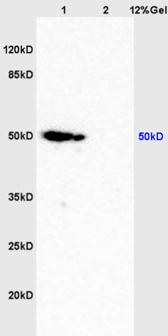 Sample:
Brain (Mouse) Lysate at 40 ug
Lung (Mouse) Lysate at 40 ug
Primary: Anti-caspase-9 p10 (SL8502R) at 1/300 dilution
Secondary: HRP conjugated Goat-Anti-rabbit IgG (SL0295G-HRP) at 1/5000 dilution
Predicted band size: 10/50 kD
Observed band size: 50 kD
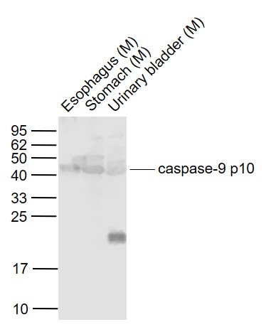 Sample:
Lane 1: Esophagus (Mouse) Lysate at 40 ug
Lane 2: Stomach (Mouse) Lysate at 40 ug
Lane 3: Urinary bladder (Mouse) Lysate at 40 ug
Primary:
Anti-caspase-9 p10 (SL8502R) at 1/1000 dilution
Secondary: IRDye800CW Goat Anti-Rabbit IgG at 1/20000 dilution
Predicted band size: 46-51/37/35/10 kD
Observed band size: 46 kD
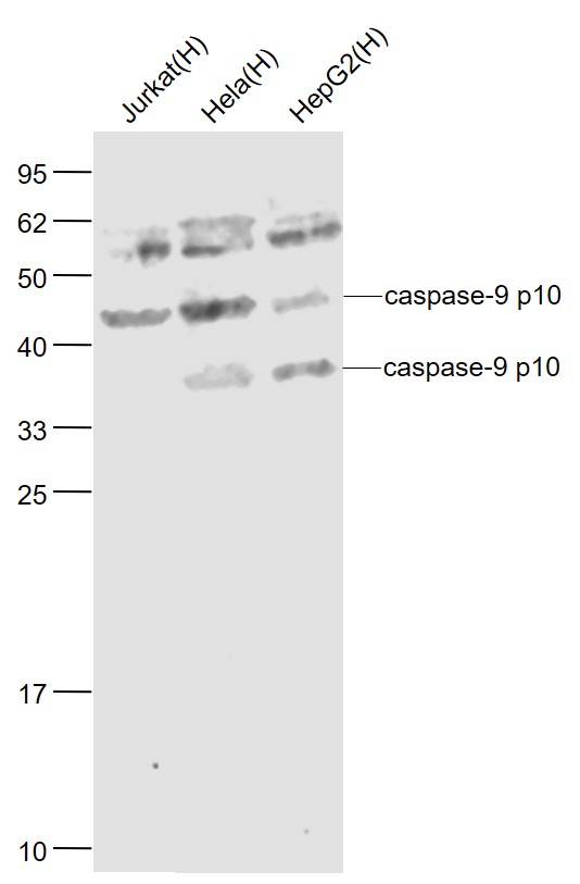 Sample:
Jurkat(Human) Cell Lysate at 30 ug
Hela(Human) Cell Lysate at 30 ug
HepG2(Human) Cell Lysate at 30 ug
Primary: Anti-caspase-9 p10 (SL8502R) at 1/1000 dilution
Secondary: IRDye800CW Goat Anti-Rabbit IgG at 1/20000 dilution
Predicted band size: 46-51/37/35/10 kD
Observed band size: 46/37 kD
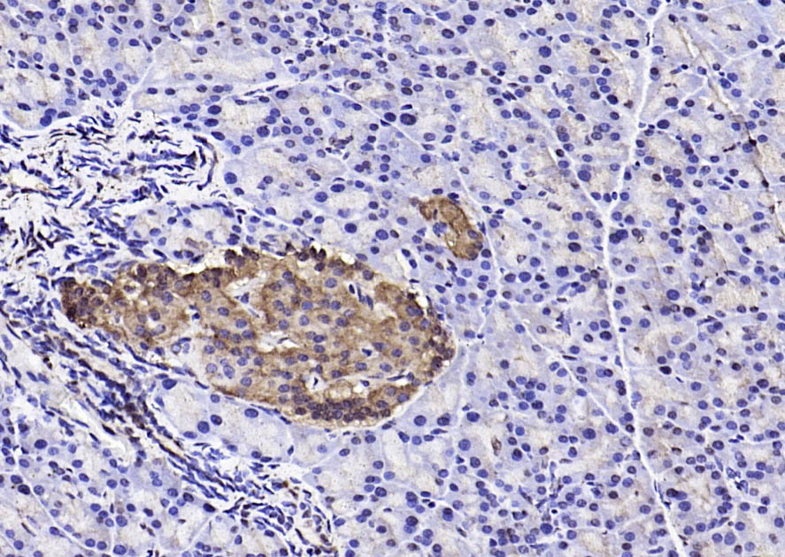 Paraformaldehyde-fixed, paraffin embedded (rat pancreas); Antigen retrieval by boiling in sodium citrate buffer (pH6.0) for 15min; Block endogenous peroxidase by 3% hydrogen peroxide for 20 minutes; Blocking buffer (normal goat serum) at 37°C for 30min; Antibody incubation with (caspase-9 p10) Polyclonal Antibody, Unconjugated (SL8502R) at 1:200 overnight at 4°C, followed by operating according to SP Kit(Rabbit) (sp-0023) instructionsand DAB staining.
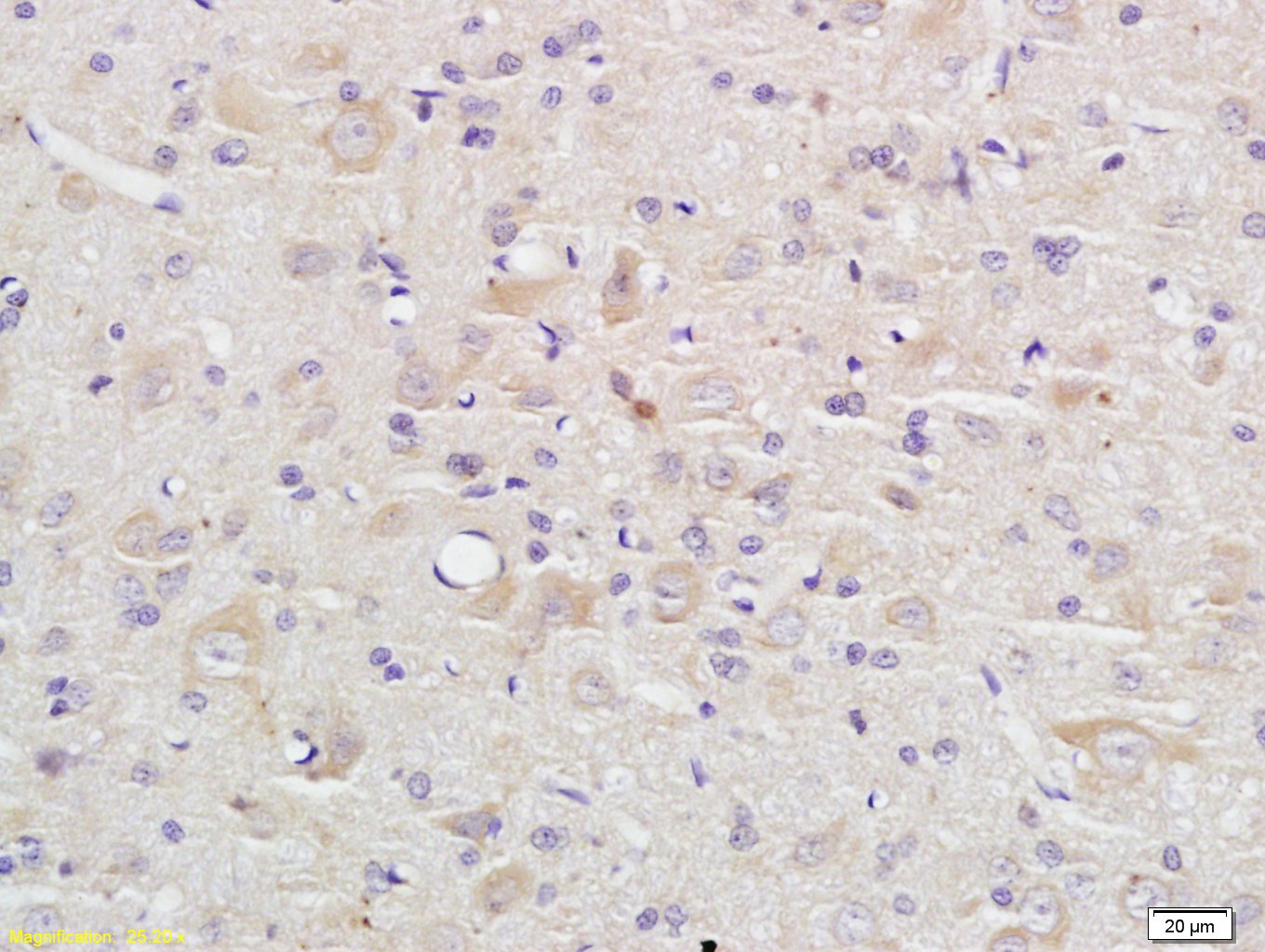 Tissue/cell: rat brain tissue; 4% Paraformaldehyde-fixed and paraffin-embedded;
Antigen retrieval: citrate buffer ( 0.01M, pH 6.0 ), Boiling bathing for 15min; Block endogenous peroxidase by 3% Hydrogen peroxide for 30min; Blocking buffer (normal goat serum,C-0005) at 37℃ for 20 min;
Incubation: Anti-caspase-9 p10 Polyclonal Antibody, Unconjugated(SL8502R) 1:200, overnight at 4°C, followed by conjugation to the secondary antibody(SP-0023) and DAB(C-0010) staining
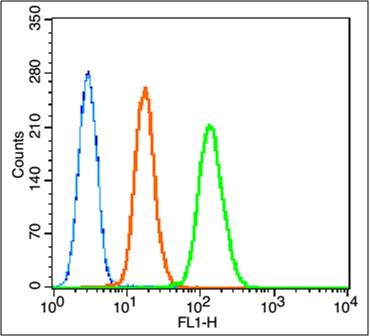 Blank control:K562 (fixed with 80% methanol (5 min) and and then permeabilized with 0.01M PBS-Tween for 20 min).
Primary Antibody:Rabbit Anti-caspase-9 p10 antibody (SL8502R,Green); Dilution: 1μg in 100 μL 1X PBS containing 0.5% BSA;
Isotype Control Antibody: Rabbit IgG(orange) ,used under the same conditions;
Secondary Antibody: Goat anti-rabbit IgG-FITC(white blue), Dilution: 1:200 in 1 X PBS containing 0.5% BSA.
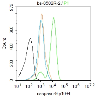 Blank control:K562.
Primary Antibody (green line): Rabbit Anti-caspase-9 p10 antibody (SL8502R)
Dilution: 2μg /10^6 cells;
Isotype Control Antibody (orange line): Rabbit IgG .
Secondary Antibody : Goat anti-rabbit IgG-FITC
Dilution: 0.5μg /test.
Protocol
The cells were fixed with 4% PFA (10min at room temperature)and then permeabilized with 90% ice-cold methanol for 20 min at-20℃. The cells were then incubated in 5%BSA to block non-specific protein-protein interactions for 30 min at room temperature .Cells stained with Primary Antibody for 30 min at room temperature. The secondary antibody used for 40 min at room temperature. Acquisition of 20,000 events was performed.
Cartpieces
Totalgoods,subtotals:¥Checkout
References (0)
No References
Bought notes(bought amounts latest0)
No one bought this product
User Comment(Total0User Comment Num)
- No comment


 +86 571 56623320
 +86 18668110335

