
Rabbit Anti-Fbxw7 antibody
AGO; Archipelago homolog; Archipelago, Drosophila, homolog of antibody CDC4; DKFZp686F23254; F box and WD 40 domain protein 7 (archipelago homolog, Drosophila); F box and WD 40 domain protein 7; F box and WD repeat domain containing 7; F box protein FBW7;
View History [Clear]
Details
Product Name Fbxw7 Chinese Name Fbxw7蛋白抗体 Alias AGO; Archipelago homolog; Archipelago, Drosophila, homolog of antibody CDC4; DKFZp686F23254; F box and WD 40 domain protein 7 (archipelago homolog, Drosophila); F box and WD 40 domain protein 7; F box and WD repeat domain containing 7; F box protein FBW7; F box protein FBX30; F box protein SEL10; F-box and WD-40 domain-containing protein 7; F-box protein FBX30; F-box/WD repeat-containing protein 7; FBW6; FBW7; FBX30; FBXO30; FBXW6; FBXW7; FBXW7_HUMAN; FLJ16457; hAgo; hCdc4; Homolog of C elegans sel 10; Homolog of C.elegans sel10; SEL-10; SEL10. literatures Research Area Tumour Cell biology immunology Neurobiology Immunogen Species Rabbit Clonality Polyclonal React Species Human, Mouse, Rat, (predicted: Dog, Pig, Cow, Horse, ) Applications ELISA=1:5000-10000 IHC-P=1:100-500 IHC-F=1:100-500 Flow-Cyt=1ug/Test IF=1:50-200 (Paraffin sections need antigen repair)
not yet tested in other applications.
optimal dilutions/concentrations should be determined by the end user.Theoretical molecular weight 78kDa Cellular localization The nucleus cytoplasmic Form Liquid Concentration 1mg/ml immunogen KLH conjugated synthetic peptide derived from human Fbxw7/CDC4: 501-600/707 Lsotype IgG Purification affinity purified by Protein A Buffer Solution 0.01M TBS(pH7.4) with 1% BSA, 0.03% Proclin300 and 50% Glycerol. Storage Shipped at 4℃. Store at -20 °C for one year. Avoid repeated freeze/thaw cycles. Attention This product as supplied is intended for research use only, not for use in human, therapeutic or diagnostic applications. PubMed PubMed Product Detail The F-box protein family is characterized by an approximately 40 amino acid motif known as the F-box. F-box proteins constitute one of the four subunits of ubiquitin protein ligase complex called SCFs (SKP1-cullin-F-box), which function in phosphorylation-dependent ubiquitination. One family member, Cdc4, also known as AGO, FBW7, FBXW7, FBX30, SEL10, and FLJ11071, maps to human chromosome 4q31.3. Alternative splicing of this gene generates four transcript variants. In addition to an F-box, Cdc4 contains seven tandem WD40 repeats. Cdc4 binds directly to cyclin E and targets cyclin E for ubiquitin-mediated degradation. Mutations of the Cdc4 gene are detected in ovarian and breast cancer cell lines, suggesting that the gene may be involved in the pathogenesis of human cancers.
Function:
Substrate recognition component of a SCF (SKP1-CUL1-F-box protein) E3 ubiquitin-protein ligase complex which mediates the ubiquitination and subsequent proteasomal degradation of target proteins. Probably recognizes and binds to phosphorylated target proteins. Involved in the degradation of cyclin-E, MYC, NOTCH1 released notch intracellular domain (NICD), and probably PSEN1.
Subunit:
Component of the SCF(FBXW7) complex consisting of CUL1, RBX1, SKP1 and FBXW7. Interacts with PSEN1, cyclin E, NOTCH1 NICD, NOTCH4 NICD and SKP1. Interacts with MYC (when phosphorylated). Isoform 1 interacts with USP28, leading to counteract ubiquitination of MYC. Isoform 4 interacts (via WD repeats) with SV40 large T antigen (via CPD region). Forms a trimeric complex with NOTCH1 and SGK1.
Subcellular Location:
Isoform 1: Nucleus, nucleoplasm. Isoform 2: Cytoplasm. Isoform 4: Nucleus, nucleolus. Nucleus.
Tissue Specificity:
Isoform 1 is widely expressed. Isoform 4 is expressed in brain.
Post-translational modifications:
Phosphorylated upon DNA damage, probably by ATM or ATR.
Similarity:
Contains 1 F-box domain.
Contains 7 WD repeats.
SWISS:
Q969H0
Gene ID:
55294
Database links:Entrez Gene: 55294 Human
Entrez Gene: 50754 Mouse
Omim: 606278 Human
SwissProt: Q969H0 Human
SwissProt: Q8VBV4 Mouse
Unigene: 561245 Human
Unigene: 196475 Mouse
Product Picture 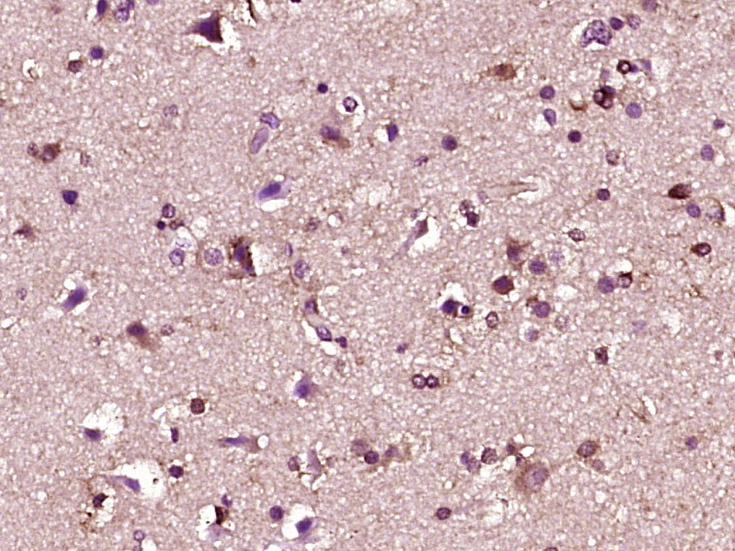 Paraformaldehyde-fixed, paraffin embedded (human brain glioma); Antigen retrieval by boiling in sodium citrate buffer (pH6.0) for 15min; Block endogenous peroxidase by 3% hydrogen peroxide for 20 minutes; Blocking buffer (normal goat serum) at 37°C for 30min; Antibody incubation with (Fbxw7) Polyclonal Antibody, Unconjugated (SL8394R) at 1:400 overnight at 4°C, followed by operating according to SP Kit(Rabbit) (sp-0023) instructionsand DAB staining.
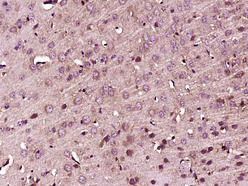 Paraformaldehyde-fixed, paraffin embedded (Mouse brain); Antigen retrieval by boiling in sodium citrate buffer (pH6.0) for 15min; Block endogenous peroxidase by 3% hydrogen peroxide for 20 minutes; Blocking buffer (normal goat serum) at 37°C for 30min; Antibody incubation with (Fbxw7) Polyclonal Antibody, Unconjugated (SL8394R) at 1:400 overnight at 4°C, followed by operating according to SP Kit(Rabbit) (sp-0023) instructionsand DAB staining.
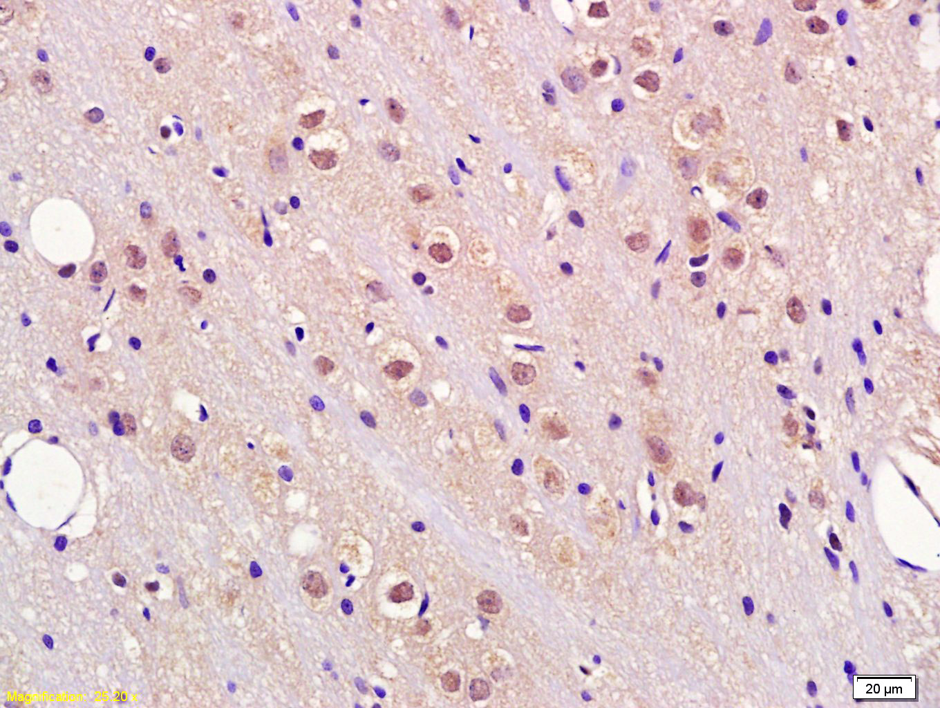 Tissue/cell: rat brain tissue; 4% Paraformaldehyde-fixed and paraffin-embedded;
Antigen retrieval: citrate buffer ( 0.01M, pH 6.0 ), Boiling bathing for 15min; Block endogenous peroxidase by 3% Hydrogen peroxide for 30min; Blocking buffer (normal goat serum,C-0005) at 37℃ for 20 min;
Incubation: Anti-Fbxw7/CDC4 Polyclonal Antibody, Unconjugated(SL8394R) 1:200, overnight at 4°C, followed by conjugation to the secondary antibody(SP-0023) and DAB(C-0010) staining
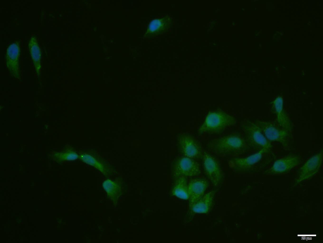 MCF-7 cell; 4% Paraformaldehyde-fixed; Triton X-100 at room temperature for 20 min; Blocking buffer (normal goat serum, C-0005) at 37°C for 20 min; Antibody incubation with (Fbxw7) polyclonal Antibody, Unconjugated (SL8394R) 1:100, 90 minutes at 37°C; followed by a conjugated Goat Anti-Rabbit IgG antibody at 37°C for 90 minutes, DAPI (blue, C02-04002) was used to stain the cell nuclei.
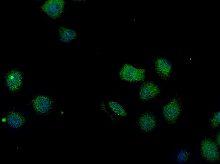 HepG2 cell; 4% Paraformaldehyde-fixed; Triton X-100 at room temperature for 20 min; Blocking buffer (normal goat serum, C-0005) at 37°C for 20 min; Antibody incubation with (Fbxw7) polyclonal Antibody, Unconjugated (SL8394R) 1:100, 90 minutes at 37°C; followed by a conjugated Goat Anti-Rabbit IgG antibody at 37°C for 90 minutes, DAPI (blue, C02-04002) was used to stain the cell nuclei.
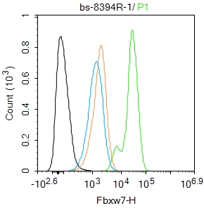 Blank control(black line):MCF-7.
Primary Antibody (green line): Rabbit Anti-Fbxw7 antibody (SL8394R)
Dilution:1ug/Test;
Secondary Antibody(white blue line): Goat anti-rabbit IgG-AF488
Dilution: 0.5ug/Test.
Isotype control(orange line): Normal Rabbit IgG
Protocol
The cells were fixed with 4% PFA (10min at room temperature)and then permeabilized with 90% ice-cold methanol for 20 min at -20℃, The cells were then incubated in 5%BSA to block non-specific protein-protein interactions for 30 min at room temperature .Cells stained with Primary Antibody for 30 min at room temperature. The secondary antibody used for 40 min at room temperature. Acquisition of 20,000 events was performed. Blank control(black line):MCF-7.
Primary Antibody (green line): Rabbit Anti-Fbxw7 antibody (SL8394R)
Dilution:1ug/Test;
Secondary Antibody(white blue line): Goat anti-rabbit IgG-AF488
Dilution: 0.5ug/Test.
Isotype control(orange line): Normal Rabbit IgG
Protocol
The cells were fixed with 4% PFA (10min at room temperature)and then permeabilized with 90% ice-cold methanol for 20 min at -20℃, The cells were then incubated in 5%BSA to block non-specific protein-protein interactions for 30 min at room temperature .Cells stained with Primary Antibody for 30 min at room temperature. The secondary antibody used for 40 min at room temperature. Acquisition of 20,000 events was performed.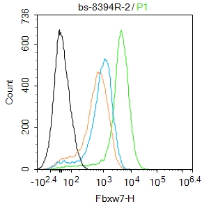 Blank control:Mouse spleen.
Primary Antibody (green line): Rabbit Anti-Fbxw7 antibody (SL8394R)
Dilution: 2μg /10^6 cells;
Isotype Control Antibody (orange line): Rabbit IgG .
Secondary Antibody : Goat anti-rabbit IgG-FITC
Dilution: 1μg /test.
Protocol
The cells were fixed with 4% PFA (10min at room temperature)and then permeabilized with 90% ice-cold methanol for 20 min at-20℃. The cells were then incubated in 5%BSA to block non-specific protein-protein interactions for 30 min at room temperature .Cells stained with Primary Antibody for 30 min at room temperature. The secondary antibody used for 40 min at room temperature. Acquisition of 20,000 events was performed.
Cartpieces
Totalgoods,subtotals:¥Checkout
References (0)
No References
Bought notes(bought amounts latest0)
No one bought this product
User Comment(Total0User Comment Num)
- No comment


 +86 571 56623320
 +86 18668110335

