Rabbit Anti-phospho-LATS1+LATS2 (Thr1079 +Thr1041)antibody
LATS1 +LATS2 (phospho T1079 + T1041); Serine/threonine protein kinase LATS2; KPM; Large tumor supressor, homolog 1; LATS, large tumor suppressor, homolog 1 (Drosophila); LATS, large tumor suppressor, homolog 2 (Drosophila); LATS1 +LATS2 (phospho T1079 + T
View History [Clear]
Details
Product Name phospho-LATS1+LATS2 (Thr1079 +Thr1041) Chinese Name 磷酸化Tumour抑制基因LATS1/LATS2抗体 Alias LATS1 +LATS2 (phospho T1079 + T1041); Serine/threonine protein kinase LATS2; KPM; Large tumor supressor, homolog 1; LATS, large tumor suppressor, homolog 1 (Drosophila); LATS, large tumor suppressor, homolog 2 (Drosophila); LATS1 +LATS2 (phospho T1079 + T1041); p-LATS1 +LATS2(Thr1079 +Thr1041); RGD1564085; Serine/threonine protein kinase LATS1; WARTS; WARTS protein kinase; wts; 4932411G09Rik; AV277261; AW208599; AW228608; FLJ13161; LATS1_HUMAN. literatures Product Type Phosphorylated anti Research Area Tumour Cell biology Signal transduction Cyclin Kinases and Phosphatases Immunogen Species Rabbit Clonality Polyclonal React Species Human, Mouse, (predicted: Rat, Chicken, Dog, Pig, Cow, Horse, Rabbit, ) Applications WB=1:500-2000 ELISA=1:5000-10000 IHC-P=1:100-500 IHC-F=1:100-500 IF=1:100-500 (Paraffin sections need antigen repair)
not yet tested in other applications.
optimal dilutions/concentrations should be determined by the end user.Theoretical molecular weight 124kDa Cellular localization cytoplasmic Form Liquid Concentration 1mg/ml immunogen KLH conjugated synthesised phosphopeptide derived from human LATS1 around the phosphorylation site of Thr1079: EF(P-T)FR Lsotype IgG Purification affinity purified by Protein A Buffer Solution 0.01M TBS(pH7.4) with 1% BSA, 0.03% Proclin300 and 50% Glycerol. Storage Shipped at 4℃. Store at -20 °C for one year. Avoid repeated freeze/thaw cycles. Attention This product as supplied is intended for research use only, not for use in human, therapeutic or diagnostic applications. PubMed PubMed Product Detail Negative regulator of YAP1 in the Hippo signaling pathway that plays a pivotal role in organ size control and tumor suppression by restricting proliferation and promoting apoptosis. The core of this pathway is composed of a kinase cascade wherein MST1/MST2, in complex with its regulatory protein SAV1, phosphorylates and activates LATS1/2 in complex with its regulatory protein MOB1, which in turn phosphorylates and inactivates YAP1 oncoprotein and WWTR1/TAZ. Phosphorylation of YAP1 by LATS2 inhibits its translocation into the nucleus to regulate cellular genes important for cell proliferation, cell death, and cell migration. Acts as a tumor suppressor which plays a critical role in centrosome duplication, maintenance of mitotic fidelity and genomic stability. Negatively regulates G1/S transition by down-regulating cyclin E/CDK2 kinase activity. Negative regulator of the androgen receptor.
Function:
Negative regulator of YAP1 in the Hippo signaling pathway that plays a pivotal role in organ size control and tumor suppression by restricting proliferation and promoting apoptosis. The core of this pathway is composed of a kinase cascade wherein STK3/MST2 and STK4/MST1, in complex with its regulatory protein SAV1, phosphorylates and activates LATS1/2 in complex with its regulatory protein MOB1, which in turn phosphorylates and inactivates YAP1 oncoprotein and WWTR1/TAZ. Phosphorylation of YAP1 by LATS1 inhibits its translocation into the nucleus to regulate cellular genes important for cell proliferation, cell death, and cell migration. Acts as a tumor suppressor which plays a critical role in maintenance of ploidy through its actions in both mitotic progression and the G1 tetraploidy checkpoint. Negatively regulates G2/M transition by down-regulating CDK1 kinase activity. Involved in the control of p53 expression. Affects cytokinesis by regulating actin polymerization through negative modulation of LIMK1. May also play a role in endocrine function.
Subunit:
Complexes with CDK1 in early mitosis. LATS1-associated CDK1 has no mitotic cyclin partner and no apparent kinase activity. Binds phosphorylated ZYX, locating this protein to the mitotic spindle and suggesting a role for actin regulatory proteins during mitosis. Binds to and colocalizes with LIMK1 at the actomyosin contractile ring during cytokinesis. Interacts (via PPxY motif 2) with YAP1 (via WW domains). Interacts with MOB1A and MOB1B. Interacts with LIMD1, WTIP and AJUBA.
Subcellular Location:
Cytoplasm, cytoskeleton, centrosome. Note=Localizes to the centrosomes throughout interphase but migrates to the mitotic apparatus, including spindle pole bodies, mitotic spindle, and midbody, during mitosis.
Tissue Specificity:
Expressed in all adult tissues examined except for lung and kidney.
Post-translational modifications:
Autophosphorylated and phosphorylated during M-phase of the cell cycle. Phosphorylated by STK3/MST2 at Ser-909 and Thr-1079, which results in its activation. Phosphorylated upon DNA damage, probably by ATM or ATR. Phosphorylation at Ser-464 by NUAK1 and NUAK2 leads to decreased protein level and is required to regulate cellular senescence and cellular ploidy.
Similarity:
Belongs to the protein kinase superfamily. AGC Ser/Thr protein kinase family.
Contains 1 AGC-kinase C-terminal domain.
Contains 1 protein kinase domain.
Contains 1 UBA domain.
SWISS:
O95835
Gene ID:
26524
Database links:Entrez Gene: 26524 Human
Entrez Gene: 9113 Human
Entrez Gene: 16798 Mouse
Entrez Gene: 50523 Mouse
Omim: 603473 Human
Omim: 604861 Human
SwissProt: O95835 Human
SwissProt: Q9NRM7 Human
SwissProt: Q7TSJ6 Mouse
SwissProt: Q8BYR2 Mouse
Product Picture
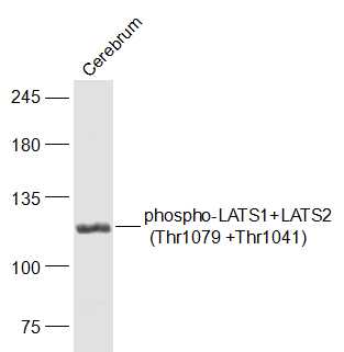
Cerebrum (Mouse) Lysate at 40 ug
Primary: Anti-phospho-LATS1+LATS2 (Thr1079 +Thr1041) (SL7913R) at 1/1000 dilution
Secondary: IRDye800CW Goat Anti-Rabbit IgG at 1/20000 dilution
Predicted band size: 124 kD
Observed band size: 124 kD
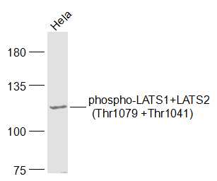
Sample:
Hela(Human) Cell Lysate at 30 ug
Primary: Anti-phospho-LATS1+LATS2 (Thr1079 +Thr1041) (SL7913R) at 1/1000 dilution
Secondary: IRDye800CW Goat Anti-Rabbit IgG at 1/20000 dilution
Predicted band size: 124 kD
Observed band size: 124 kD
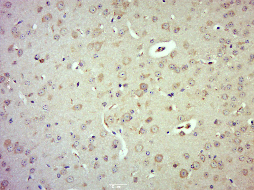
Paraformaldehyde-fixed, paraffin embedded (Mouse brain); Antigen retrieval by boiling in sodium citrate buffer (pH6.0) for 15min; Block endogenous peroxidase by 3% hydrogen peroxide for 20 minutes; Blocking buffer (normal goat serum) at 37°C for 30min; Antibody incubation with (phospho-LATS1+LATS2 (Thr1079 +Thr1041)) Polyclonal Antibody, Unconjugated (SL7913R) at 1:500 overnight at 4°C, followed by a conjugated secondary (sp-0023) for 20 minutes and DAB staining.
Bought notes(bought amounts latest0)
No one bought this product
User Comment(Total0User Comment Num)
- No comment


 +86 571 56623320
 +86 18668110335

