
Rabbit Anti-NEDD4 antibody
Cell proliferation-inducing gene 53 protein; E3 ubiquitin protein ligase Nedd4; E3 ubiquitin-protein ligase NEDD4; KIAA0093; NEDD 4; NEDD-4; Nedd4; NEDD4_HUMAN; Neural precursor cell expressed developmentally down regulated 4; Neural precursor cell expres
View History [Clear]
Details
Product Name NEDD4 Chinese Name 神经前体细胞发育下调蛋白4抗体 Alias Cell proliferation-inducing gene 53 protein; E3 ubiquitin protein ligase Nedd4; E3 ubiquitin-protein ligase NEDD4; KIAA0093; NEDD 4; NEDD-4; Nedd4; NEDD4_HUMAN; Neural precursor cell expressed developmentally down regulated 4; Neural precursor cell expressed developmentally down-regulated protein 4. literatures Research Area Cell biology Neurobiology Signal transduction Cyclin Cell differentiation Immunogen Species Rabbit Clonality Polyclonal React Species Human, Rat, (predicted: Mouse, Chicken, Dog, Pig, Cow, Horse, Rabbit, ) Applications WB=1:500-2000 ELISA=1:5000-10000 IHC-P=1:100-500 IHC-F=1:100-500 Flow-Cyt=3ug/test ICC=1:100-500 IF=1:100-500 (Paraffin sections need antigen repair)
not yet tested in other applications.
optimal dilutions/concentrations should be determined by the end user.Theoretical molecular weight 145kDa Cellular localization cytoplasmic The cell membrane Form Liquid Concentration 1mg/ml immunogen KLH conjugated synthetic peptide derived from human SRD5A2: 801-900/1319 Lsotype IgG Purification affinity purified by Protein A Buffer Solution 0.01M TBS(pH7.4) with 1% BSA, 0.03% Proclin300 and 50% Glycerol. Storage Shipped at 4℃. Store at -20 °C for one year. Avoid repeated freeze/thaw cycles. Attention This product as supplied is intended for research use only, not for use in human, therapeutic or diagnostic applications. PubMed PubMed Product Detail E3 ubiquitin-protein ligase which accepts ubiquitin from an E2 ubiquitin-conjugating enzyme in the form of a thioester and then directly transfers the ubiquitin to targeted substrates. Involved in the pathway leading to the degradation of VEGFR-2/KDFR, independently of its ubiquitin-ligase activity. Monoubiquitinates IGF1R at multiple sites, thus leading to receptor internalization and degradation in lysosomes. According to PubMed:18562292 the direct link between NEDD4 and PTEN regulation through polyubiquitination described in PubMed:17218260 is questionable. Involved in ubiquitination of ERBB4 intracellular domain E4ICD. Involved in the budding of many viruses. Part of a signaling complex composed of NEDD4, RAP2A and TNIK which regulates neuronal dendrite extension and arborization during development. Ubiquitinates TNK2 and regulates EGF-induced degradation of EGFR and TNF2.
Function:
E3 ubiquitin-protein ligase which accepts ubiquitin from an E2 ubiquitin-conjugating enzyme in the form of a thioester and then directly transfers the ubiquitin to targeted substrates. Involved in the pathway leading to the degradation of VEGFR-2/KDFR, independently of its ubiquitin-ligase activity. Monoubiquitinates IGF1R at multiple sites, thus leading to receptor internalization and degradation in lysosomes. Ubiquitinates FGFR1, leading to receptor internalization and degradation in lysosomes. According to PubMed:18562292 the direct link between NEDD4 and PTEN regulation through polyubiquitination described in PubMed:17218260 is questionable. Involved in ubiquitination of ERBB4 intracellular domain E4ICD. Involved in the budding of many viruses. Part of a signaling complex composed of NEDD4, RAP2A and TNIK which regulates neuronal dendrite extension and arborization during development. Ubiquitinates TNK2 and regulates EGF-induced degradation of EGFR and TNF2.
Subunit:
Interacts with UBE2D2. Binds SCNN1A, SCNN1B and SCNN1G. Binds, in vitro, through the WW2 and WW3 domains, to neural isoforms of ENAH that contain the PPSY motif. Interacts with BEAN1, LITAF, RNF11, WBP1, WBP2, TMEPAI and PRRG2 (By similarity). Interacts with NDFIP1 and NDFIP2; this interaction activates the E3 ubiquitin-protein ligase and may induce its recruitment to exosomes. Interaction with PTEN is questionable according to PubMed:18562292. Interacts with viral proteins that contain a late- budding motif P-P-P-Y. This interaction is essential for viral particle budding of a lot of retroviruses, like HTLV-1 Gag and MLV Gag. Interacts (via C2 domain) with GRB10 (via SH2 domain). Interacts with ERBB4. Interacts with TNIK; the interaction is direct, allows the TNIK-dependent recruitment of RAP2A and its ubiquitination by NEDD4. Interacts (via WW3 domain) with TNK2; EGF promotes this interaction. Interacts (via WW3 domain) with FGFR1 (via C-terminus).
Subcellular Location:
Cytoplasm. Cell membrane; Peripheral membrane protein. Note=Recruited to the plasma membrane by GRB10. Once complexed with GRB10 and IGF1R, follows IGF1R internalization, remaining associated with early endosomes. Uncouples from IGF1R-containing endosomes before the sorting of the receptor to the lysosomal compartment. May be recruited to exosomes by NDFIP1..
Post-translational modifications:
Auto-ubiquitinated.
Similarity:
Contains 1 HECT (E6AP-type E3 ubiquitin-protein ligase) domain.
Contains 4 WW domains.
SWISS:
P46934
Gene ID:
4734
Database links:Entrez Gene: 4734 Human
Entrez Gene: 17999 Mouse
Omim: 602278 Human
SwissProt: P46934 Human
SwissProt: P46935 Mouse
Unigene: 1565 Human
Unigene: 279923 Mouse
Unigene: 99540 Rat
Product Picture 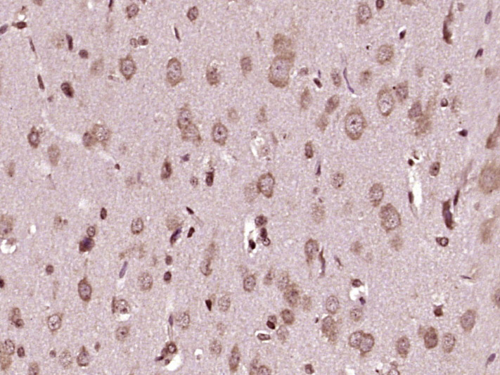 Paraformaldehyde-fixed, paraffin embedded (rat brain tissue); Antigen retrieval by boiling in sodium citrate buffer (pH6.0) for 15min; Block endogenous peroxidase by 3% hydrogen peroxide for 20 minutes; Blocking buffer (normal goat serum) at 37°C for 30min; Antibody incubation with (NEDD4) Polyclonal Antibody, Unconjugated (SL7877R) at 1:400 overnight at 4°C, followed by operating according to SP Kit(Rabbit) (sp-0023) instructionsand DAB staining.
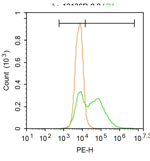 Blank control:A549.
Primary Antibody (green line): Rabbit Anti-NEDD4 antibody (SL7877R)
Dilution: 1μg /10^6 cells;
Isotype Control Antibody (orange line): Rabbit IgG .
Secondary Antibody : Goat anti-rabbit IgG-PE
Dilution: 3μg /test.
Protocol
The cells were fixed with 4% PFA (10min at room temperature)and then permeabilized with 20% PBST for 20 min at room temperature. The cells were then incubated in 5% BSA to block non-specific protein-protein interactions for 30 min at at room temperature .Cells stained with Primary Antibody for 30 min at room temperature. The secondary antibody used for 40 min at room temperature. Acquisition of 20,000 events was performed.
Cartpieces
Totalgoods,subtotals:¥Checkout
References (0)
No References
Bought notes(bought amounts latest0)
No one bought this product
User Comment(Total0User Comment Num)
- No comment


 +86 571 56623320
 +86 18668110335

