
Rabbit Anti-EDG3 antibody
EDG 3; EDG3; Endothelial differentiation G protein coupled receptor 3; Endothelial differentiation G-protein coupled receptor 3; Endothelial differentiation gene 3; Endothelial differentiation sphingolipid G protein coupled receptor 3; FLJ37523; FLJ93220;
View History [Clear]
Details
Product Name EDG3 Chinese Name 内皮分化型G protein-coupled receptor3抗体 Alias EDG 3; EDG3; Endothelial differentiation G protein coupled receptor 3; Endothelial differentiation G-protein coupled receptor 3; Endothelial differentiation gene 3; Endothelial differentiation sphingolipid G protein coupled receptor 3; FLJ37523; FLJ93220; G protein coupled receptor endothelial differentiation gene 3; LPB 3; LPB3; S1P receptor 3; S1P receptor Edg 3; S1P receptor Edg-3; S1P receptor EDG3; S1P3; S1PR3; S1PR3_HUMAN; Sphingosine 1 phosphate receptor 3; Sphingosine 1 phosphate receptor Edg 3; Sphingosine 1 phosphate receptor Edg3; Sphingosine 1-phosphate receptor 3; Sphingosine 1-phosphate receptor Edg-3. literatures Research Area Cell biology Signal transduction transcriptional regulatory factor G protein-coupled receptor G protein signal Immunogen Species Rabbit Clonality Polyclonal React Species Human, Mouse, Rat, (predicted: Cow, Rabbit, ) Applications WB=1:500-2000 ELISA=1:5000-10000 IHC-P=1:100-500 IHC-F=1:100-500 Flow-Cyt=0.2u/test IF=1:100-500 (Paraffin sections need antigen repair)
not yet tested in other applications.
optimal dilutions/concentrations should be determined by the end user.Theoretical molecular weight 42kDa Cellular localization The cell membrane Form Liquid Concentration 1mg/ml immunogen KLH conjugated synthetic peptide derived from human EDG3: 145-250/378 <Extracellular> Lsotype IgG Purification affinity purified by Protein A Buffer Solution 0.01M TBS(pH7.4) with 1% BSA, 0.03% Proclin300 and 50% Glycerol. Storage Shipped at 4℃. Store at -20 °C for one year. Avoid repeated freeze/thaw cycles. Attention This product as supplied is intended for research use only, not for use in human, therapeutic or diagnostic applications. PubMed PubMed Product Detail This gene encodes a member of the EDG family of receptors, which are G protein-coupled receptors. This protein has been identified as a functional receptor for sphingosine 1-phosphate and likely contributes to the regulation of angiogenesis and vascular endothelial cell function. [provided by RefSeq, Jul 2008].
Function:
Receptor for the lysosphingolipid sphingosine 1-phosphate (S1P). S1P is a bioactive lysophospholipid that elicits diverse physiological effect on most types of cells and tissues. When expressed in rat HTC4 hepatoma cells, is capable of mediating S1P-induced cell proliferation and suppression of apoptosis.
Subcellular Location:
Cell membrane; Multi-pass membrane protein.
Tissue Specificity:
Expressed in all tissues, but most abundantly in heart, placenta, kidney, and liver.
Similarity:
Belongs to the G-protein coupled receptor 1 family.
SWISS:
Q99500
Gene ID:
1903
Database links:Entrez Gene: 1903 Human
Entrez Gene: 13610 Mouse
Omim: 601965 Human
SwissProt: Q99500 Human
SwissProt: Q9Z0U9 Mouse
Unigene: 585118 Human
Unigene: 136736 Mouse
Unigene: 23918 Rat
Product Picture 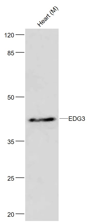 Sample:
Lane1: Heart (Mouse) Lysate at 40 ug
Primary: Anti-EDG3 (SL7541R) at 1/300 dilution
Secondary: HRP conjugated Goat-Anti-rabbit IgG (SL0295G-HRP) at 1/5000 dilution
Predicted band size: 42 kD
Observed band size: 46 kD
 Sample:
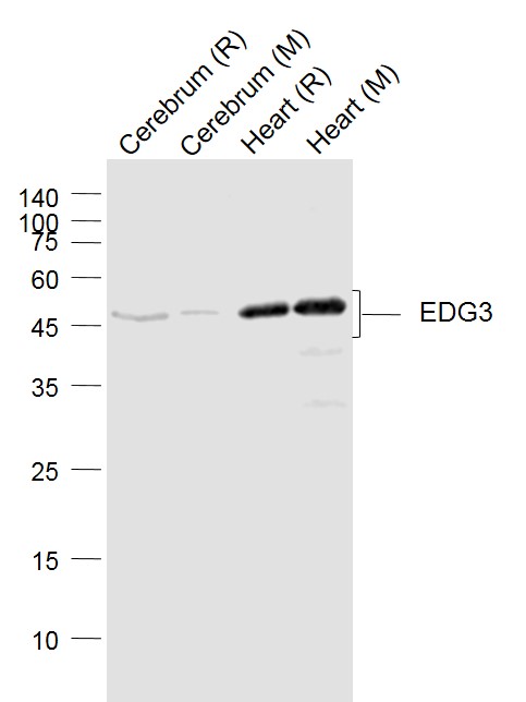
Lane 1: Cerebrum (Rat) Lysate at 40 ug
Lane 2: Cerebrum (Mouse) Lysate at 40 ug
Lane 3: Heart (Rat) Lysate at 40 ug
Lane 4: Heart (Mouse) Lysate at 40 ug
Primary: Anti-EDG3 (SL7541R) at 1/1000 dilution
Secondary: IRDye800CW Goat Anti-Rabbit IgG at 1/20000 dilution
Predicted band size: 46 kD
Observed band size: 46 kD
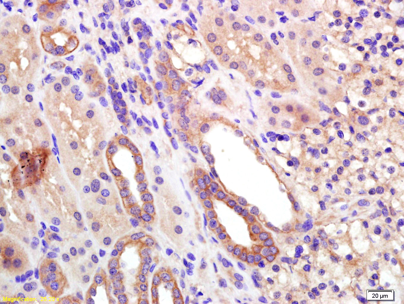 Tissue/cell: human kidney tissue; 4% Paraformaldehyde-fixed and paraffin-embedded;
Antigen retrieval: citrate buffer ( 0.01M, pH 6.0 ), Boiling bathing for 15min; Block endogenous peroxidase by 3% Hydrogen peroxide for 30min; Blocking buffer (normal goat serum,C-0005) at 37∩ for 20 min;
Incubation: Anti-EDG3 Polyclonal Antibody, Unconjugated(SL7541R) 1:200, overnight at 4∑C, followed by conjugation to the secondary antibody(SP-0023) and DAB(C-0010) staining
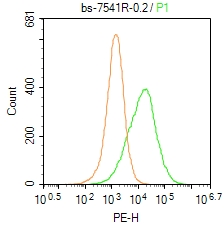 Blank control: Hela.
Primary Antibody (green line): Rabbit Anti-EDG3 antibody (SL7541R)
Dilution: 0.2μg /10^6 cells;
Isotype Control Antibody (orange line): Rabbit IgG .
Secondary Antibody : Goat anti-rabbit IgG-PE
Dilution: 1μg /test.
Protocol
The cells were incubated in 5%BSA to block non-specific protein-protein interactions for 30 min at at room temperature .Cells stained with Primary Antibody for 30 min at room temperature. The secondary antibody used for 40 min at room temperature. Acquisition of 20,000 events was performed.
Cartpieces
Totalgoods,subtotals:¥Checkout
References (0)
No References
Bought notes(bought amounts latest0)
No one bought this product
User Comment(Total0User Comment Num)
- No comment


 +86 571 56623320
 +86 18668110335

