
Rabbit Anti-Melan A antibody
MAR1_HUMAN; Melanoma antigen recognized by T-cells 1; MLANA; MART1; Antigen LB39-AA; Antigen SK29-AA; Protein Melan-A;
View History [Clear]
Details
Product Name Melan A Chinese Name 黑色素瘤相关抗原/黑色素-A抗体 Alias MAR1_HUMAN; Melanoma antigen recognized by T-cells 1; MLANA; MART1; Antigen LB39-AA; Antigen SK29-AA; Protein Melan-A; Research Area Tumour Cell biology immunology t-lymphocyte Immunogen Species Rabbit Clonality Polyclonal React Species Human, Mouse, (predicted: Rat, ) Applications WB=1:500-2000 ELISA=1:5000-10000 IHC-P=1:100-500 IHC-F=1:100-500 ICC=1:100-500 IF=1:100-500 (Paraffin sections need antigen repair)
not yet tested in other applications.
optimal dilutions/concentrations should be determined by the end user.Theoretical molecular weight 13kDa Cellular localization cytoplasmic Form Liquid Concentration 1mg/ml immunogen KLH conjugated synthetic peptide derived from mouse Melan A: 1-80/113 Lsotype IgG Purification affinity purified by Protein A Buffer Solution 0.01M TBS(pH7.4) with 1% BSA, 0.03% Proclin300 and 50% Glycerol. Storage Shipped at 4℃. Store at -20 °C for one year. Avoid repeated freeze/thaw cycles. Attention This product as supplied is intended for research use only, not for use in human, therapeutic or diagnostic applications. PubMed PubMed Product Detail Melanoma-associated antigens recognized by cytotoxic T lymphocytes (CTL) have been grouped into three categories: melanocyte differentiation antigens, cancer/testis-specific antigens and mutated or aberrantly expressed antigens. Many of these antigens consist of peptides that are presented to T cells by HLA molecules; they represent potential targets for cancer immunotherapy. Melan-A (also designated MART-1) is a melanocyte differentiation antigen that is specific to melanomas, melanocyte cell lines and retina. Melan-A peptide is recognized by most HLA-A2-restricted tumor-specific tumor-infiltrating lymphocytes in patients with melanoma. Antimelanoma cytotoxic T lymphocytes can be generated with a Melan-A peptide, implicating Melan-A as a potential candidate for antigen-specific immunotherapy in melanoma patients.
Function:
Involved in melanosome biogenesis by ensuring the stability of GPR143. Plays a vital role in the expression, stability, trafficking, and processing of melanocyte protein PMEL, which is critical to the formation of stage II melanosomes.
Subunit:
Interacts with PMEL. Interacts with GPR143.
Subcellular Location:
Endoplasmic reticulum membrane. Golgi apparatus. Golgi apparatus > trans-Golgi network membrane. Melanosome. Also found in small vesicles and tubules dispersed over the entire cytoplasm. A small fraction of the protein is inserted into the membrane in an inverted orientation. Inversion of membrane topology results in the relocalization of the protein from a predominant Endoplasmic reticulum membrane. Golgi apparatus. Golgi apparatus > trans-Golgi network membrane. Melanosome. Also found in small vesicles and tubules dispersed over the entire cytoplasm. A small fraction of the protein is inserted into the membrane in an inverted orientation. Inversion of membrane topology results in the relocalization of the protein from a predominant Golgi/post-Golgi area to the endoplasmic reticulum. Melanoma cells expressing the protein with an inverted membrane topology are more effectively recognized by specific cytolytic T-lymphocytes than those expressing the protein in its native membrane orientation.
Tissue Specificity:
Expression is restricted to melanoma and melanocyte cell lines and retina.
Post-translational modifications:
Acylated.
SWISS:
Q2TA50
Gene ID:
77836
Database links:Entrez Gene: 77836 Mouse
Product Picture 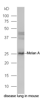 Protein: disease lung in mouse lysate;
Primary: rabbit Anti-Melan A (SL7362R) at 1:300;
Secondary: HRP conjugated Goat-Anti-rabbit IgG(SL0295G-HRP) at 1: 5000;
Predicted band size: 26 kD
Observed band size: 26 kD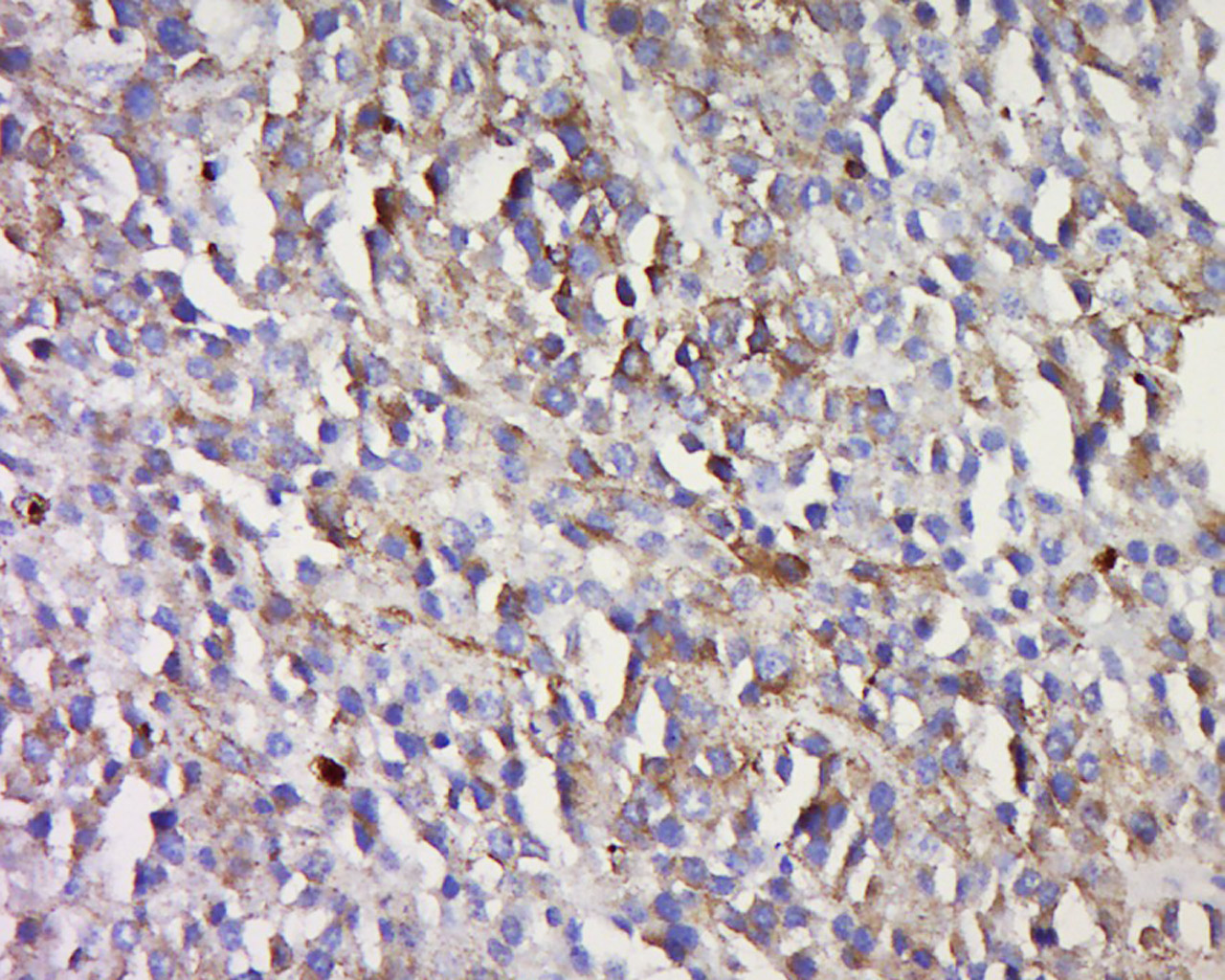 Tissue/cell: human melanoma tissue; 4% Paraformaldehyde-fixed and paraffin-embedded;
Antigen retrieval: citrate buffer ( 0.01M, pH 6.0 ), Boiling bathing for 15min; Block endogenous peroxidase by 3% Hydrogen peroxide for 30min; Blocking buffer (normal goat serum,C-0005) at 37℃ for 20 min;
Incubation: Anti-Melan A Polyclonal Antibody, Unconjugated(SL7362R) 1:500, overnight at 4°C, followed by conjugation to the secondary antibody(SP-0023) and DAB(C-0010) staining
Cartpieces
Totalgoods,subtotals:¥Checkout
References (0)
No References
Bought notes(bought amounts latest0)
No one bought this product
User Comment(Total0User Comment Num)
- No comment


 +86 571 56623320
 +86 18668110335

