
Rabbit Anti-WT1 antibody
WT1_HUMAN; Wilms tumor protein; WT33; WT1 transcription factor; GUD; AWT1; WAGR; WT-1; WT33; NPHS4; WIT-2;
View History [Clear]
Details
Product Name WT1 Chinese Name 肾母细胞瘤蛋白抗体 Alias WT1_HUMAN; Wilms tumor protein; WT33; WT1 transcription factor; GUD; AWT1; WAGR; WT-1; WT33; NPHS4; WIT-2; literatures Research Area Tumour Cell biology immunology Developmental biology TumourCell biologyMaker Epigenetics Immunogen Species Rabbit Clonality Polyclonal React Species Human, Mouse, Rat, (predicted: Chicken, Dog, Pig, Cow, Sheep, ) Applications WB=1:500-2000 ELISA=1:5000-10000 IHC-P=1:100-500 IHC-F=1:100-500 Flow-Cyt=1ug/test IF=1:100-500 (Paraffin sections need antigen repair)
not yet tested in other applications.
optimal dilutions/concentrations should be determined by the end user.Theoretical molecular weight 55kDa Cellular localization The nucleus cytoplasmic Form Liquid Concentration 1mg/ml immunogen KLH conjugated synthetic peptide derived from human WT1: 301-400/449 Lsotype IgG Purification affinity purified by Protein A Buffer Solution 0.01M TBS(pH7.4) with 1% BSA, 0.03% Proclin300 and 50% Glycerol. Storage Shipped at 4℃. Store at -20 °C for one year. Avoid repeated freeze/thaw cycles. Attention This product as supplied is intended for research use only, not for use in human, therapeutic or diagnostic applications. PubMed PubMed Product Detail Transcription factor that plays an important role in cellular development and cell survival. Regulates the expression of numerous target genes, including EPO. Plays an essential role for development of the urogenital system. Recognizes and binds to the DNA sequence 5'-CGCCCCCGC-3'. It has a tumor suppressor as well as an oncogenic role in tumor formation. Function may be isoform-specific: isoforms lacking the KTS motif may act as transcription factors. Isoforms containing the KTS motif may bind mRNA and play a role in mRNA metabolism or splicing. Isoform 1 has lower affinity for DNA, and can bind RNA.
Function:
Transcription factor that plays an important role in cellular development and cell survival. Regulates the expression of numerous target genes, including EPO. Plays an essential role for development of the urogenital system. Recognizes and binds to the DNA sequence 5'-CGCCCCCGC-3'. It has a tumor suppressor as well as an oncogenic role in tumor formation. Function may be isoform-specific: isoforms lacking the KTS motif may act as transcription factors. Isoforms containing the KTS motif may bind mRNA and play a role in mRNA metabolism or splicing. Isoform 1 has lower affinity for DNA, and can bind RNA.
Subunit:
Homodimer. Interacts with WTIP. Interacts with actively translating polysomes. Detected in nuclear ribonucleoprotein (mRNP) particles. Interacts with HNRNPU via the zinc-finger region. Interacts with U2AF2. Interacts with CITED2. Interacts with ZNF224 via the zinc-finger region. Interacts with WTAP and SRY. Interacts with FAM123B/WTX. Interacts with RBM4.
Subcellular Location:
Nucleus. Nucleus, nucleolus. Cytoplasm. Note=Shuttles between nucleus and cytoplasm.
Isoform 1: Nucleus speckle.
Isoform 4: Nucleus, nucleoplasm.
Tissue Specificity:
Expressed in the kidney and a subset of hematopoietic cells.
DISEASE:
Defects in WT1 are the cause of Frasier syndrome (FS) [MIM:136680]. FS is characterized by a slowly progressing nephropathy leading to renal failure in adolescence or early adulthood, male pseudohermaphroditism, and no Wilms tumor. As for histological findings of the kidneys, focal glomerular sclerosis is often observed. There is phenotypic overlap with Denys-Drash syndrome. Inheritance is autosomal dominant.
Defects in WT1 are the cause of Wilms tumor 1 (WT1) [MIM:194070]. WT is an embryonal malignancy of the kidney that affects approximately 1 in 10'000 infants and young children. It occurs both in sporadic and hereditary forms.
Defects in WT1 are the cause of Denys-Drash syndrome (DDS) [MIM:194080]. DDS is a typical nephropathy characterized by diffuse mesangial sclerosis, genital abnormalities, and/or Wilms tumor. There is phenotypic overlap with WAGR syndrome and Frasier syndrome. Inheritance is autosomal dominant, but most cases are sporadic.
Similarity:
Belongs to the EGR C2H2-type zinc-finger protein family.
Contains 4 C2H2-type zinc fingers.
SWISS:
P19544
Gene ID:
7490
Database links:Entrez Gene: 7490 Human
Entrez Gene: 22431 Mouse
Omim: 607102 Human
SwissProt: P19544 Human
SwissProt: P22561 Mouse
Unigene: 591980 Human
Unigene: 389339 Mouse
Unigene: 92531 Rat
Product Picture 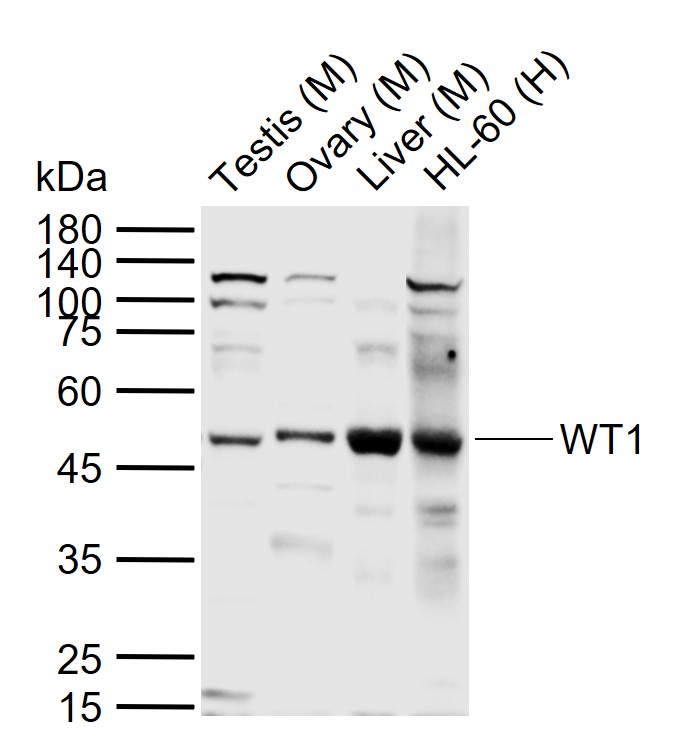 Sample:
Lane 1: Mouse Testis tissue lysates
Lane 2: Mouse Ovary tissue lysates
Lane 3: Mouse Liver tissue lysates
Lane 4: Human HL-60 cell lysates
Primary: Anti-WT1 (SL6983R) at 1/1000 dilution
Secondary: IRDye800CW Goat Anti-Rabbit IgG at 1/20000 dilution
Predicted band size: 55 kDa
Observed band size: 50 kDa
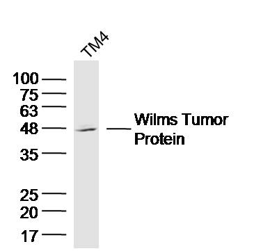 Sample: TM4 Cell (Mouse) Lysate at 40 ug
Primary: Anti-Wilms Tumor Protein (SL6983R) at 1/300 dilution
Secondary: IRDye800CW Goat Anti-Rabbit IgG at 1/20000 dilution
Predicted band size: 55 kD
Observed band size: 48 kD
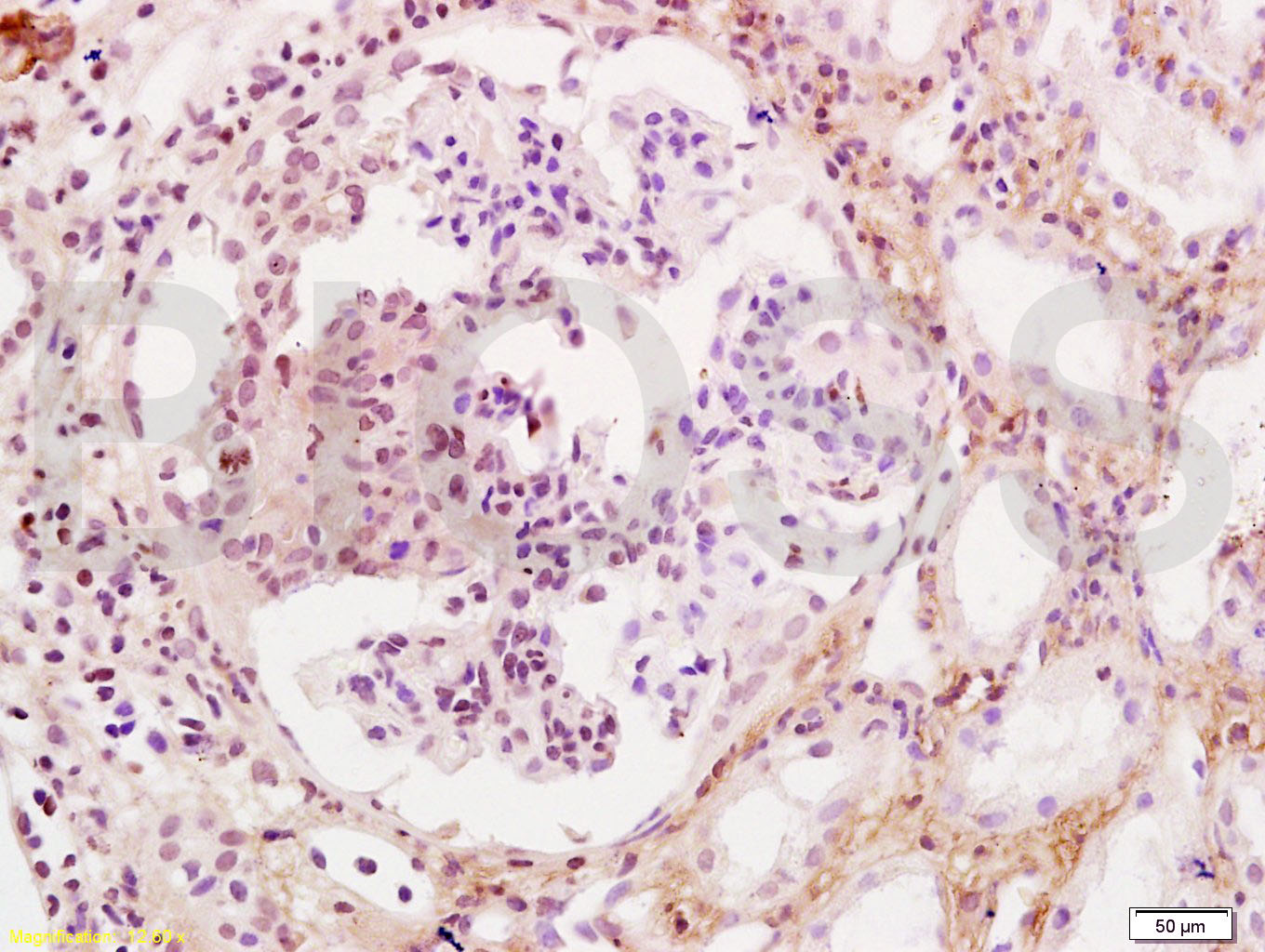 Tissue/cell: human kidney tissue; 4% Paraformaldehyde-fixed and paraffin-embedded;
Antigen retrieval: citrate buffer ( 0.01M, pH 6.0 ), Boiling bathing for 15min; Block endogenous peroxidase by 3% Hydrogen peroxide for 30min; Blocking buffer (normal goat serum,C-0005) at 37℃ for 20 min;
Incubation: Anti-WT-1/Wilms Tumor Protein Polyclonal Antibody, Unconjugated(SL6983R) 1:200, overnight at 4°C, followed by conjugation to the secondary antibody(SP-0023) and DAB(C-0010) staining
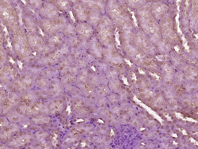 Paraformaldehyde-fixed, paraffin embedded (Mouse kidney); Antigen retrieval by boiling in sodium citrate buffer (pH6.0) for 15min; Block endogenous peroxidase by 3% hydrogen peroxide for 20 minutes; Blocking buffer (normal goat serum) at 37°C for 30min; Antibody incubation with (Wilms Tumor Protein) Polyclonal Antibody, Unconjugated (SL6983R) at 1:400 overnight at 4°C, followed by operating according to SP Kit(Rabbit) (sp-0023) instructionsand DAB staining.
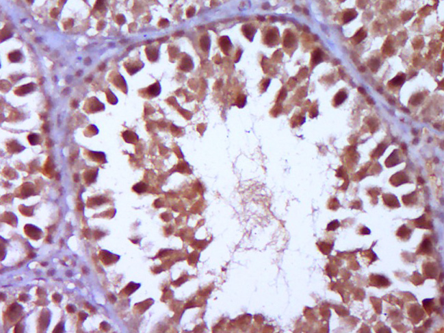 Paraformaldehyde-fixed, paraffin embedded (Rat testis); Antigen retrieval by boiling in sodium citrate buffer (pH6.0) for 15min; Block endogenous peroxidase by 3% hydrogen peroxide for 20 minutes; Blocking buffer (normal goat serum) at 37°C for 30min; Antibody incubation with (Wilms Tumor Protein) Polyclonal Antibody, Unconjugated (SL6983R) at 1:400 overnight at 4°C, followed by operating according to SP Kit(Rabbit) (sp-0023) instructionsand DAB staining.
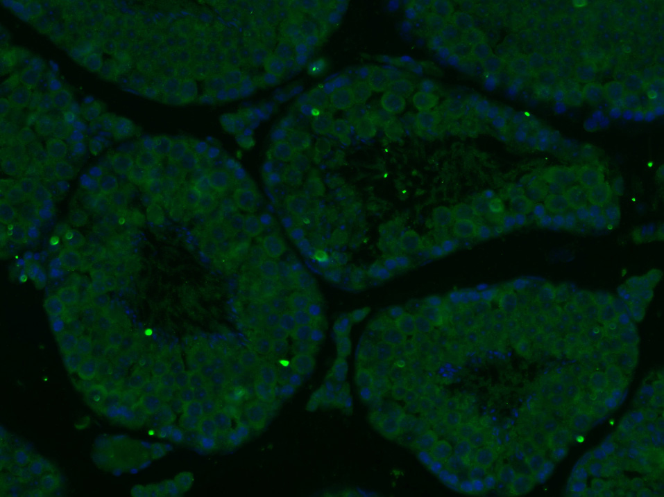 Paraformaldehyde-fixed, paraffin embedded (Mouse testis); Antigen retrieval by boiling in sodium citrate buffer (pH6.0) for 15min; Block endogenous peroxidase by 3% hydrogen peroxide for 20 minutes; Blocking buffer (normal goat serum) at 37°C for 30min; Antibody incubation with (Wilms Tumor Protein) Polyclonal Antibody, Unconjugated (SL6983R) at 1:400 overnight at 4°C, followed by a conjugated Goat Anti-Rabbit IgG antibody (SL0295G-FITC) for 90 minutes, and DAPI for nuclei staining.
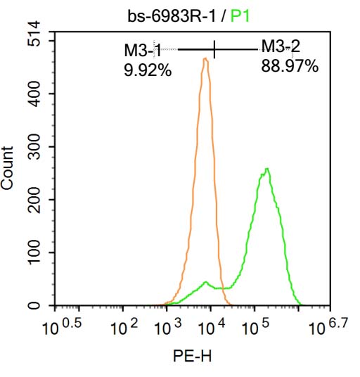 Blank control:Molt-4.
Primary Antibody (green line): Rabbit Anti-Wilms Tumor Protein antibody (SL6983R)
Dilution: 1μg /10^6 cells;
Isotype Control Antibody (orange line): Rabbit IgG .
Secondary Antibody : Goat anti-rabbit IgG-AF647
Dilution: 1μg /test.
Protocol
The cells were fixed with 4% PFA (10min at room temperature)and then permeabilized with 90% ice-cold methanol for 20 min at-20℃. The cells were then incubated in 5%BSA to block non-specific protein-protein interactions for 30 min at at room temperature .Cells stained with Primary Antibody for 30 min at room temperature. The secondary antibody used for 40 min at room temperature. Acquisition of 20,000 events was performed.
Cartpieces
Totalgoods,subtotals:¥Checkout
References (0)
No References
Bought notes(bought amounts latest0)
No one bought this product
User Comment(Total0User Comment Num)
- No comment


 +86 571 56623320
 +86 18668110335

