
Rabbit Anti-CD95/FAS antibody
Apo-1; ALPS 1A; ALPS1A; APO 1; Apo 1 antigen; APO 1 cell surface antigen; Apo-1 antigen; APO1; Apo1 antigen; APO1 cell surface antigen; Apoptosis antigen 1; Apoptosis mediating surface antigen FAS; Apoptosis-mediating surface antigen FAS; APT 1; APT1; CD
View History [Clear]
Details
Product Name CD95/FAS Chinese Name 载Lipoprotein1抗体 Alias Apo-1; ALPS 1A; ALPS1A; APO 1; Apo 1 antigen; APO 1 cell surface antigen; Apo-1 antigen; APO1; Apo1 antigen; APO1 cell surface antigen; Apoptosis antigen 1; Apoptosis mediating surface antigen FAS; Apoptosis-mediating surface antigen FAS; APT 1; APT1; CD 95; CD 95 antigen; CD95; CD95 antigen; Delta Fas; Delta Fas/APO 1/CD95; Delta Fas/APO1/CD95; FAS 1; FAS 827dupA; Fas AMA; FAS; FAS Antigen; FAS1; FASLG receptor; FASTM; TNF receptor superfamily, member 6; TNFRSF 6; TNFRSF6; TNR6_HUMAN; Tumor necrosis factor receptor superfamily member 6. literatures Research Area Cell biology immunology Apoptosis Immunogen Species Rabbit Clonality Polyclonal React Species Human, Mouse, Rat, (predicted: Pig, ) Applications WB=1:500-2000 ELISA=1:5000-10000 Flow-Cyt=2μg/Test
not yet tested in other applications.
optimal dilutions/concentrations should be determined by the end user.Theoretical molecular weight 34kDa Cellular localization The cell membrane Secretory protein Form Liquid Concentration 1mg/ml immunogen KLH conjugated synthetic peptide derived from human FAS/Apo-1/CD95: 81-170/335 <Extracellular> Lsotype IgG Purification affinity purified by Protein A Buffer Solution 0.01M TBS(pH7.4) with 1% BSA, 0.03% Proclin300 and 50% Glycerol. Storage Shipped at 4℃. Store at -20 °C for one year. Avoid repeated freeze/thaw cycles. Attention This product as supplied is intended for research use only, not for use in human, therapeutic or diagnostic applications. PubMed PubMed Product Detail The protein encoded by this gene is a member of the TNF-receptor superfamily. This receptor contains a death domain. It has been shown to play a central role in the physiological regulation of programmed cell death, and has been implicated in the pathogenesis of various malignancies and diseases of the immune system. The interaction of this receptor with its ligand allows the formation of a death-inducing signaling complex that includes Fas-associated death domain protein (FADD), caspase 8, and caspase 10. The autoproteolytic processing of the caspases in the complex triggers a downstream caspase cascade, and leads to apoptosis. This receptor has been also shown to activate NF-kappaB, MAPK3/ERK1, and MAPK8/JNK, and is found to be involved in transducing the proliferating signals in normal diploid fibroblast and T cells. Several alternatively spliced transcript variants have been described, some of which are candidates for nonsense-mediated mRNA decay (NMD). The isoforms lacking the transmembrane domain may negatively regulate the apoptosis mediated by the full length isoform. [provided by RefSeq, Mar 2011]
Function:
Receptor for TNFSF6/FASLG. The adapter molecule FADD recruits caspase-8 to the activated receptor. The resulting death-inducing signaling complex (DISC) performs caspase-8 proteolytic activation which initiates the subsequent cascade of caspases (aspartate-specific cysteine proteases) mediating apoptosis. FAS-mediated apoptosis may have a role in the induction of peripheral tolerance, in the antigen-stimulated suicide of mature T-cells, or both. The secreted isoforms 2 to 6 block apoptosis (in vitro).
Subunit:
Binds DAXX. Interacts with HIPK3. Part of a complex containing HIPK3 and FADD. Binds RIPK1 and FAIM2. Interacts with BRE and FEM1B. Interacts with FADD.
Subcellular Location:
Isoform 1: Cell membrane; Single-pass type I membrane protein. Isoform 2, 3, 4, 5, 6: Secreted.
Tissue Specificity:
Isoform 1 and isoform 6 are expressed at equal levels in resting peripheral blood mononuclear cells. After activation there is an increase in isoform 1 and decrease in the levels of isoform 6.
Post-translational modifications:
N- and O-glycosylated. O-glycosylated with core 1 or possibly core 8 glycans.
DISEASE:
Defects in FAS are the cause of autoimmune lymphoproliferative syndrome type 1A (ALPS1A) [MIM:601859]; also known as Canale-Smith syndrome (CSS). ALPS is a childhood syndrome involving hemolytic anemia and thrombocytopenia with massive lymphadenopathy and splenomegaly.
Similarity:
Contains 1 death domain.
Contains 3 TNFR-Cys repeats.
SWISS:
P25445
Gene ID:
355
Database links:Entrez Gene: 355 Human
Entrez Gene: 14102 Mouse
Omim: 134637 Human
SwissProt: P25445 Human
SwissProt: P25446 Mouse
Unigene: 244139 Human
Unigene: 1626 Mouse
Unigene: 162521 Rat
Product Picture 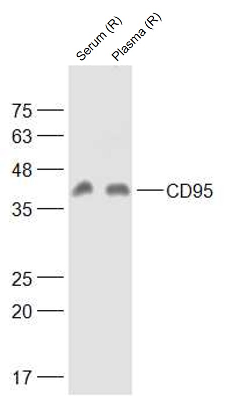 Sample:
serum (Rat) at 40 ug
plasma (Rat) at 40 ug
Primary: Anti-CD95 (SL6477R) at 1/1000 dilution
Secondary: IRDye800CW Goat Anti-Rabbit IgG at 1/20000 dilution
Predicted band size: 34 kD
Observed band size: 42 kD
 Sample:
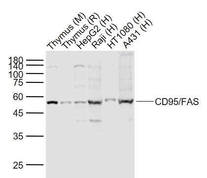
Lane 1: Thymus (Mouse) Lysate at 40 ug
Lane 2: Thymus (Rat) Lysate at 40 ug
Lane 3: HepG2 (Human) Cell Lysate at 30 ug
Lane 4: Raji (Human) Cell Lysate at 30 ug
Lane 5: HT1080 (Human) Cell Lysate at 30 ug
Lane 6: A431 (Human) Cell Lysate at 30 ug
Primary: Anti- CD95/FAS (SL6477R) at 1/1000 dilution
Secondary: IRDye800CW Goat Anti-Rabbit IgG at 1/20000 dilution
Predicted band size: 45/52 kD
Observed band size: 52 kD
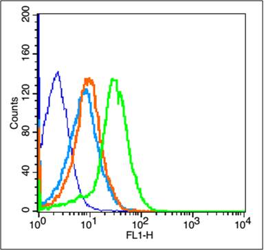 Blank control(blue):Mouse Kidney (fixed with 2% paraformaldehyde for 10 min at 37℃).
Primary Antibody:Rabbit Anti-CD95/FAS antibody (SL6477R,Green); Dilution: 1μg in 100 μL 1X PBS containing 0.5% BSA;
Isotype Control Antibody: Rabbit IgG(orange) ,used under the same conditions;
Secondary Antibody: Goat anti-rabbit IgG-FITC(white blue), Dilution: 1:200 in 1 X PBS containing 0.5% BSA.
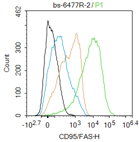 Blank control:Raji.
Primary Antibody (green line): Rabbit Anti-CD95/FAS antibody (SL6477R)
Dilution: 2ug/Test;
Secondary Antibody : Goat anti-rabbit IgG-AF488
Dilution: 0.5ug/Test.
Protocol
The cells were incubated in 5%BSA to block non-specific protein-protein interactions for 30 min at room temperature .Cells stained with Primary Antibody for 30 min at room temperature. The secondary antibody used for 40 min at room temperature. Acquisition of 20,000 events was performed.
Cartpieces
Totalgoods,subtotals:¥Checkout
References (0)
No References
Bought notes(bought amounts latest0)
No one bought this product
User Comment(Total0User Comment Num)
- No comment


 +86 571 56623320
 +86 18668110335

