
Rabbit Anti-IRAK1 antibody
Il1rak; Il1rak; Interleukin 1 receptor associated kinase 1; Interleukin 1 receptor associated kinase 1; Interleukin 1 receptor associated kinase 2; Interleukin-1 receptor-associated kinase 1; IRAK; IRAK-1; IRAK1; IRAK1; IRAK1_HUMAN; IRAK2; IRAK2; mPLK; mP
View History [Clear]
Details
Product Name IRAK1 Chinese Name 白介素-1受体相关激酶1抗体 Alias Il1rak; Il1rak; Interleukin 1 receptor associated kinase 1; Interleukin 1 receptor associated kinase 1; Interleukin 1 receptor associated kinase 2; Interleukin-1 receptor-associated kinase 1; IRAK; IRAK-1; IRAK1; IRAK1; IRAK1_HUMAN; IRAK2; IRAK2; mPLK; mPLK; Pelle; Pelle; Pelle homolog; Pelle-like protein kinase; Plpk. Research Area Cell biology immunology Signal transduction Kinases and Phosphatases Immunogen Species Rabbit Clonality Polyclonal React Species Human, Rat, (predicted: Mouse, Chicken, Dog, Pig, Cow, Rabbit, ) Applications ELISA=1:5000-10000 IHC-P=1:100-500 IHC-F=1:100-500 Flow-Cyt=1ug/Test ICC=1:100 IF=1:50-200 (Paraffin sections need antigen repair)
not yet tested in other applications.
optimal dilutions/concentrations should be determined by the end user.Theoretical molecular weight 78kDa Cellular localization The nucleus cytoplasmic Form Liquid Concentration 1mg/ml immunogen KLH conjugated synthetic peptide derived from human IRAK1: 301-400/712 Lsotype IgG Purification affinity purified by Protein A Buffer Solution 0.01M TBS(pH7.4) with 1% BSA, 0.03% Proclin300 and 50% Glycerol. Storage Shipped at 4℃. Store at -20 °C for one year. Avoid repeated freeze/thaw cycles. Attention This product as supplied is intended for research use only, not for use in human, therapeutic or diagnostic applications. PubMed PubMed Product Detail Binds to the IL-1 type I receptor following IL-1 engagement, triggering intracellular signaling cascades leading to transcriptional up-regulation and mRNA stabilization. Isoform 1 binds rapidly but is then degraded allowing isoform 2 to mediate a slower, more sustained response to the cytokine. Isoform 2 is inactive suggesting that the kinase activity of this enzyme is not required for IL-1 signaling. Once phosphorylated, IRAK1 recruits the adapter protein PELI1.
Function:
Serine/threonine-protein kinase that plays a critical role in initiating innate immune response against foreign pathogens. Involved in Toll-like receptor (TLR) and IL-1R signaling pathways. Is rapidly recruited by MYD88 to the receptor-signaling complex upon TLR activation. Association with MYD88 leads to IRAK1 phosphorylation by IRAK4 and subsequent autophosphorylation and kinase activation. Phosphorylates E3 ubiquitin ligases Pellino proteins (PELI1, PELI2 and PELI3) to promote pellino-mediated polyubiquitination of IRAK1. Then, the ubiquitin-binding domain of IKBKG/NEMO binds to polyubiquitinated IRAK1 bringing together the IRAK1-MAP3K7/TAK1-TRAF6 complex and the NEMO-IKKA-IKKB complex. In turn, MAP3K7/TAK1 activates IKKs (CHUK/IKKA and IKBKB/IKKB) leading to NF-kappa-B nuclear translocation and activation. Alternatively, phosphorylates TIRAP to promote its ubiquitination and subsequent degradation. Phosphorylates the interferon regulatory factor 7 (IRF7) to induce its activation and translocation to the nucleus, resulting in transcriptional activation of type I IFN genes, which drive the cell in an antiviral state. When sumoylated, translocates to the nucleus and phosphorylates STAT3
Subunit:
Homodimer. Interacts with TOLLIP; this interaction occurs in the cytosol prior to receptor activation. Interacts with MYD88; this interaction recruits IRAK1 to the stimulated receptor complex. Interacts with IL1RL1. Interacts with IRAK1BP1. Associates with TRAF6, PELI1 and IRAK4; this complex recruits MAP3K7/TAK1, TAB1 and TAB2 to mediate NF-kappa-B activation. Interacts (when polyubiquitinated) with IKBKG/NEMO.
Subcellular Location:
Cytoplasm. Nucleus. Note=Translocates to the nucleus when sumoylated.
Tissue Specificity:
Isoform 1 and isoform 2 are ubiquitously expressed in all tissues examined, with isoform 1 being more strongly expressed than isoform 2.
Post-translational modifications:
Following recruitment on the activated receptor complex, phosphorylated on Thr-209, probably by IRAK4, resulting in a conformational change of the kinase domain, allowing further phosphorylations to take place. Thr-387 phosphorylation in the activation loop is required to achieve full enzymatic activity.
Polyubiquitinated after cell stimulation with IL-1-beta by PELI1, PELI2 and PELI3. Polyubiquitination occurs with polyubiquitin chains linked through 'Lys-63'. Ubiquitination promotes interaction with NEMO/IKBKG. Also sumoylated; leading to nuclear translocation.
Similarity:
Belongs to the protein kinase superfamily. TKL Ser/Thr protein kinase family. Pelle subfamily.
Contains 1 death domain.
Contains 1 protein kinase domain.
SWISS:
Q62406
Gene ID:
16179
Database links:
Entrez Gene: 3654 Human
Entrez Gene: 16179 Mouse
Omim: 300283 Human
SwissProt: P51617 Human
SwissProt: Q62406 Mouse
Unigene: 522819 Human
Unigene: 38241 Mouse
Unigene: 22238 Rat
Product Picture 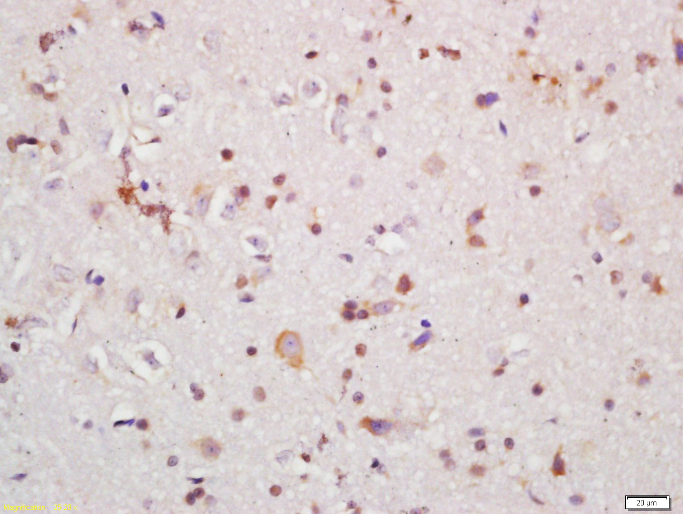 Tissue/cell: rat brain tissue; 4% Paraformaldehyde-fixed and paraffin-embedded;
Antigen retrieval: citrate buffer ( 0.01M, pH 6.0 ), Boiling bathing for 15min; Block endogenous peroxidase by 3% Hydrogen peroxide for 30min; Blocking buffer (normal goat serum,C-0005) at 37℃ for 20 min;
Incubation: Anti-IRAK1 Polyclonal Antibody, Unconjugated(SL6464R) 1:200, overnight at 4°C, followed by conjugation to the secondary antibody(SP-0023) and DAB(C-0010) staining
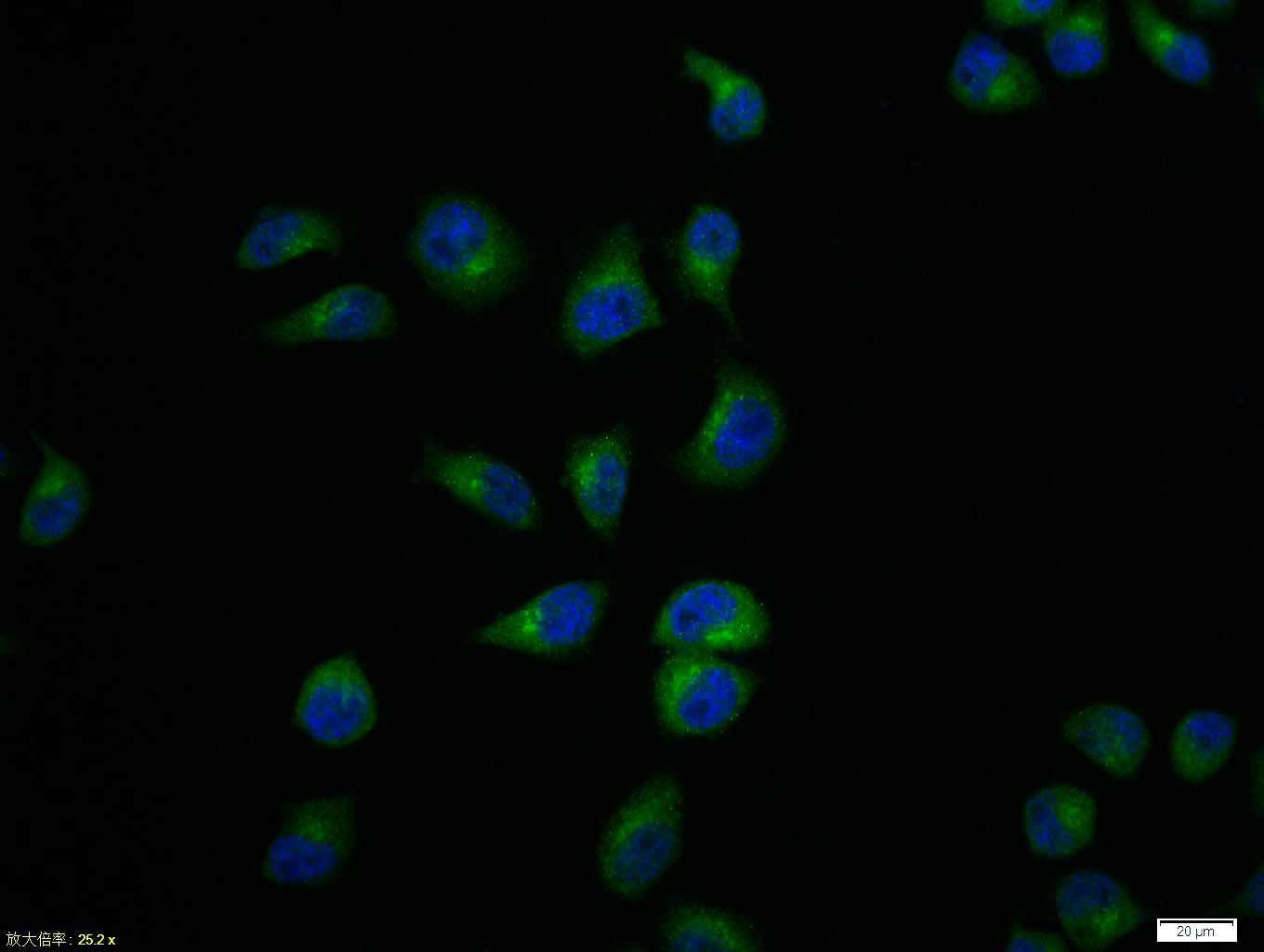 Hela cell; 4% Paraformaldehyde-fixed; Triton X-100 at room temperature for 20 min; Blocking buffer (normal goat serum, C-0005) at 37°C for 20 min; Antibody incubation with (IRAK1) polyclonal Antibody, Unconjugated (SL6464R) 1:100, 90 minutes at 37°C; followed by a conjugated Goat Anti-Rabbit IgG antibody at 37°C for 90 minutes, DAPI (blue, C02-04002) was used to stain the cell nuclei.
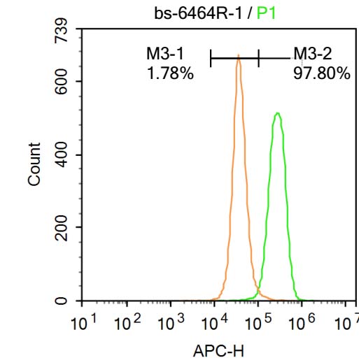 Blank control: A431.
Primary Antibody (green line): Rabbit Anti-IRAK1 antibody (SL6464R)
Dilution: 1μg /10^6 cells;
Isotype Control Antibody (orange line): Rabbit IgG .
Secondary Antibody : Goat anti-rabbit IgG-AF647
Dilution: 1μg /test.
Protocol
The cells were fixed with 4% PFA (10min at room temperature)and then permeabilized with 90% ice-cold methanol for 20 min at-20℃. The cells were then incubated in 5%BSA to block non-specific protein-protein interactions for 30 min at at room temperature .Cells stained with Primary Antibody for 30 min at room temperature. The secondary antibody used for 40 min at room temperature. Acquisition of 20,000 events was performed.
Cartpieces
Totalgoods,subtotals:¥Checkout
Bought notes(bought amounts latest0)
No one bought this product
User Comment(Total0User Comment Num)
- No comment


 +86 571 56623320


