
Rabbit Anti-IL-1 Beta antibody
IL1B_HUMAN; Interleukin-1 beta; interleukin 1 beta; IL1F2; IL-1 beta; Catabolin; IL-1; IL1F2; IL1beta; IL1-BETA; IL-1beta; IL-1 Beta; IL-1β; IL1β; IL-1B; IL1B;
View History [Clear]
Details
Product Name IL-1 Beta Chinese Name 白介素1β抗体 Alias IL1B_HUMAN; Interleukin-1 beta; interleukin 1 beta; IL1F2; IL-1 beta; Catabolin; IL-1; IL1F2; IL1beta; IL1-BETA; IL-1beta; IL-1 Beta; IL-1β; IL1β; IL-1B; IL1B; literatures Research Area Cell biology immunology Growth factors and hormones glycoprotein Immunogen Species Rabbit Clonality Polyclonal React Species Human, Mouse, Rat, (predicted: Dog, Horse, Rabbit, ) Applications WB=1:500-2000 ELISA=1:5000-10000 IHC-P=1:100-500 IHC-F=1:100-500 Flow-Cyt=2ug/Test IF=1:100-500 (Paraffin sections need antigen repair)
not yet tested in other applications.
optimal dilutions/concentrations should be determined by the end user.Theoretical molecular weight 17/32kDa Cellular localization Secretory protein Form Liquid Concentration 1mg/ml immunogen KLH conjugated synthetic peptide derived from human IL-1 Beta: 161-269/269 Lsotype IgG Purification affinity purified by Protein A Buffer Solution 0.01M TBS(pH7.4) with 1% BSA, 0.03% Proclin300 and 50% Glycerol. Storage Shipped at 4℃. Store at -20 °C for one year. Avoid repeated freeze/thaw cycles. Attention This product as supplied is intended for research use only, not for use in human, therapeutic or diagnostic applications. PubMed PubMed Product Detail The protein encoded by this gene is a member of the interleukin 1 cytokine family. This cytokine is produced by activated macrophages as a proprotein, which is proteolytically processed to its active form by caspase 1 (CASP1/ICE). This cytokine is an important mediator of the inflammatory response, and is involved in a variety of cellular activities, including cell proliferation, differentiation, and apoptosis. The induction of cyclooxygenase-2 (PTGS2/COX2) by this cytokine in the central nervous system (CNS) is found to contribute to inflammatory pain hypersensitivity. This gene and eight other interleukin 1 family genes form a cytokine gene cluster on chromosome 2. [provided by RefSeq, Jul 2008].
Function:
Produced by activated macrophages, IL-1 stimulates thymocyte proliferation by inducing IL-2 release, B-cell maturation and proliferation, and fibroblast growth factor activity. IL-1 proteins are involved in the inflammatory response, being identified as endogenous pyrogens, and are reported to stimulate the release of prostaglandin and collagenase from synovial cells.
Subunit:
Monomer.
Subcellular Location:
Secreted. Note=The lack of a specific hydrophobic segment in the precursor sequence suggests that IL-1 is released by damaged cells or is secreted by a mechanism differing from that used for other secretory proteins.
Similarity:
Belongs to the IL-1 family.
SWISS:
P01584
Gene ID:
3553
Database links:Entrez Gene: 3553 Human
Entrez Gene: 16176 Mouse
Omim: 147720 Human
SwissProt: P01584 Human
SwissProt: P10749 Mouse
Unigene: 126256 Human
Unigene: 222830 Mouse
Unigene: 9869 Rat
Product Picture 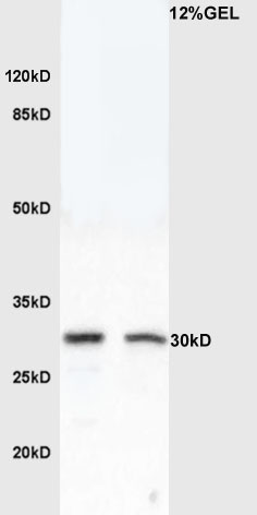 Sample:
Brain (Mouse) Lysate at 40 ug
Intestine (Mouse) Lysate at 40 ug
Primary: Anti-IL-1 Beta (SL6319R) at 1/300 dilution
Secondary: HRP conjugated Goat-Anti-rabbit IgG (SL0295G-HRP) at 1/5000 dilution
Predicted band size: 17/30 kD
Observed band size: 30 kD
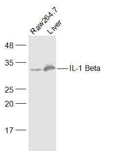 Sample:
Raw264.7(Mouse) Cell Lysate at 30 ug
Liver (Mouse) Lysate at 40 ug
Primary: Anti-IL-1 Beta (SL6319R) at 1/500 dilution
Secondary: IRDye800CW Goat Anti-Rabbit IgG at 1/20000 dilution
Predicted band size: 17/30 kD
Observed band size: 30 kD
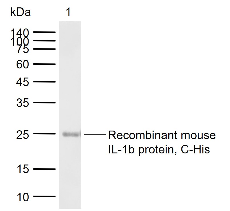 Sample:
Lane 1: Recombinant mouse IL-1b protein, C-His
Primary: Anti-IL-1 Beta (SL6319R) at 1/1000 dilution
Secondary: IRDye800CW Goat Anti-Rabbit IgG at 1/20000 dilution
Predicted band size: 17/32 kDa
Observed band size: 25 kDa
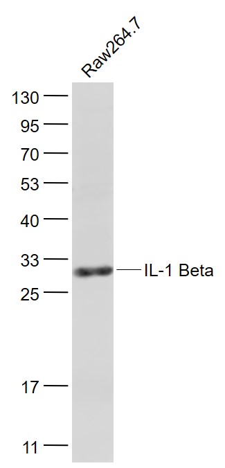 Sample:
Raw264.7(Mouse) Cell Lysate at 30 ug
Primary: Anti- IL-1 Beta (SL6319R) at 1/1000 dilution
Secondary: IRDye800CW Goat Anti-Rabbit IgG at 1/20000 dilution
Predicted band size: 17/30 kD
Observed band size: 30 kD
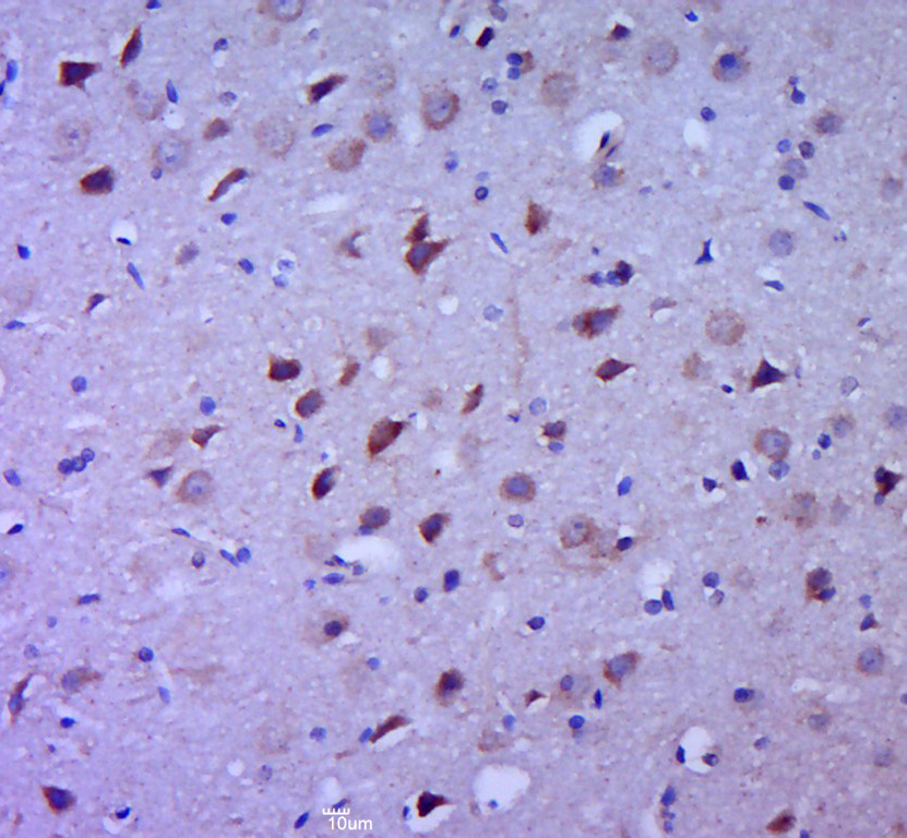 Paraformaldehyde-fixed, paraffin embedded (rat brain tissue); Antigen retrieval by boiling in sodium citrate buffer (pH6.0) for 15min; Block endogenous peroxidase by 3% hydrogen peroxide for 20 minutes; Blocking buffer (normal goat serum) at 37°C for 30min; Antibody incubation with (IL-1 Beta) Polyclonal Antibody, Unconjugated (SL6319R) at 1:400 overnight at 4°C, followed by a conjugated secondary (sp-0023) for 20 minutes and DAB staining.
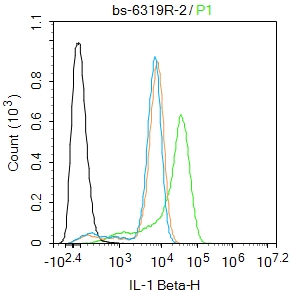 Blank control: THP-1.
Primary Antibody (green line): Rabbit Anti-IL-1 Beta antibody (SL6319R)
Dilution: 2μg /10^6 cells;
Isotype Control Antibody (orange line): Rabbit IgG .
Secondary Antibody : Goat anti-rabbit IgG-FITC
Dilution: 0.5μg /test.
Protocol
The cells were fixed with 4% PFA (10min at room temperature)and then permeabilized with 0.1% PBST for 20 min at room temperature. The cells were then incubated in 5%BSA to block non-specific protein-protein interactions for 30 min at room temperature .Cells stained with Primary Antibody for 30 min at room temperature. The secondary antibody used for 40 min at room temperature. Acquisition of 20,000 events was performed.
Cartpieces
Totalgoods,subtotals:¥Checkout
References (0)
No References
Bought notes(bought amounts latest0)
No one bought this product
User Comment(Total0User Comment Num)
- No comment


 +86 571 56623320
 +86 18668110335

