
Rabbit Anti-Prostaglandin E Receptor EP1 antibody
Prostaglandin E Receptor EP1; EP1; PGE receptor EP1 subtype; PGE2 receptor EP1 subtype; Prostaglandin E receptor 1 subtype EP1 42kDa; Prostaglandin E receptor 1 subtype EP1; Prostaglandin E2 receptor EP1 subtype; Prostanoid EP1 receptor; PE2R1_HUMAN.
View History [Clear]
Details
Product Name Prostaglandin E Receptor EP1 Chinese Name 前列腺素EP1受体抗体 Alias Prostaglandin E Receptor EP1; EP1; PGE receptor EP1 subtype; PGE2 receptor EP1 subtype; Prostaglandin E receptor 1 subtype EP1 42kDa; Prostaglandin E receptor 1 subtype EP1; Prostaglandin E2 receptor EP1 subtype; Prostanoid EP1 receptor; PE2R1_HUMAN. literatures Research Area Cell biology Signal transduction transcriptional regulatory factor G protein-coupled receptor G protein signal Immunogen Species Rabbit Clonality Polyclonal React Species Mouse, Rat, (predicted: Human, Dog, ) Applications WB=1:500-2000 ELISA=1:5000-10000 IHC-P=1:100-500 IHC-F=1:100-500 Flow-Cyt=1ug/Test IF=1:100-500 (Paraffin sections need antigen repair)
not yet tested in other applications.
optimal dilutions/concentrations should be determined by the end user.Theoretical molecular weight 42kDa Cellular localization The cell membrane Form Liquid Concentration 1mg/ml immunogen KLH conjugated synthetic peptide derived from human Prostaglandin E Receptor EP1: 61-160/402 <Extracellular> Lsotype IgG Purification affinity purified by Protein A Buffer Solution 0.01M TBS(pH7.4) with 1% BSA, 0.03% Proclin300 and 50% Glycerol. Storage Shipped at 4℃. Store at -20 °C for one year. Avoid repeated freeze/thaw cycles. Attention This product as supplied is intended for research use only, not for use in human, therapeutic or diagnostic applications. PubMed PubMed Product Detail PTGER1 is a subtype 1 receptor for prostaglandin E2 (PGE2). This receptor is coupled to the phosphatidylinositol-calcium second messenger system by G(q) proteins. PTGER1 may be an important modulator of renal function and is implicated in the smooth muscle contractile response to PGE2 in various tissues.
Subcellular Location:
Cell membrane; Multi-pass membrane protein.
Tissue Specificity:
Abundant in kidney. Lower level expression in lung, skeletal muscle and spleen, lowest expression in testis and not detected in liver brain and heart.
Similarity:
Belongs to the G-protein coupled receptor 1 family.
SWISS:
P34995
Gene ID:
5731
Database links:Entrez Gene: 5731 Human
Entrez Gene: 19216 Mouse
Omim: 176802 Human
SwissProt: P34995 Human
SwissProt: P35375 Mouse
Unigene: 159360 Human
Unigene: 474430 Mouse
Unigene: 11423 Rat
Product Picture 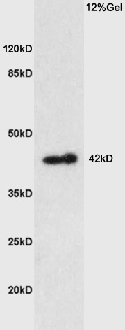 Sample: Brain (Rat) Lysate at 40 ug
Primary: Anti-Prostaglandin E Receptor EP1 (SL6316R) at 1/300 dilution
Secondary: HRP conjugated Goat-Anti-rabbit IgG (SL0295G-HRP) at 1/5000 dilution
Predicted band size: 42 kD
Observed band size: 42 kD
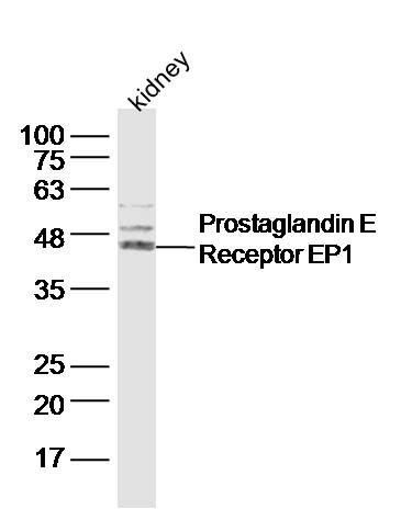 Sample: Kidney (Mouse) Lysate at 40 ug
Primary: Anti-Prostaglandin E Receptor EP1 (SL6316R) at 1/300 dilution
Secondary: IRDye800CW Goat Anti-Rabbit IgG at 1/20000 dilution
Predicted band size: 42 kD
Observed band size: 45 kD
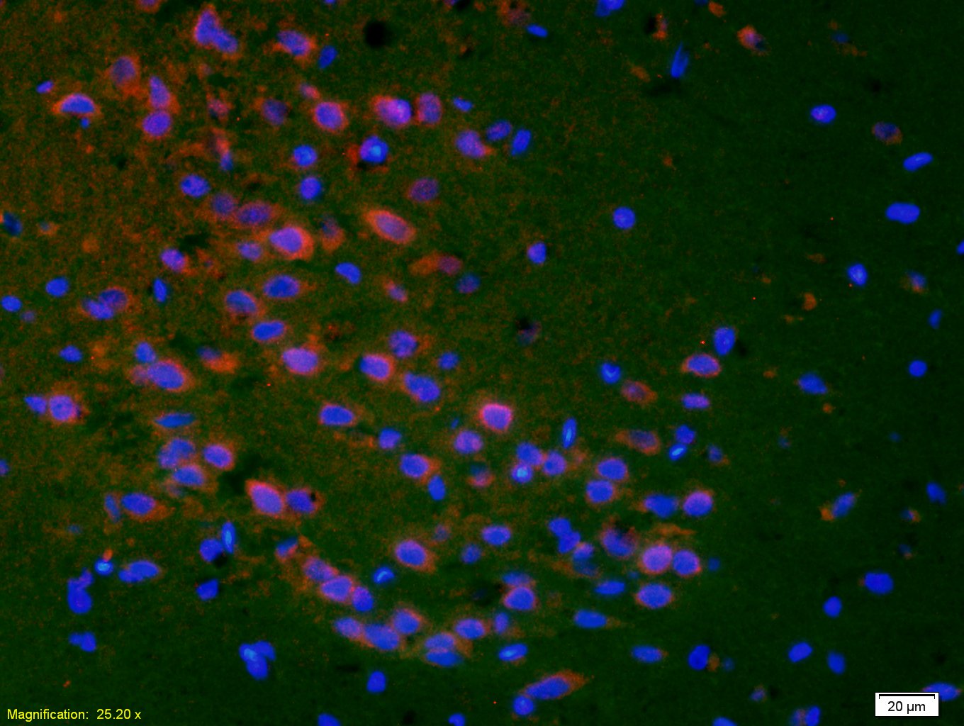 Tissue/cell: rat brain tissue;4% Paraformaldehyde-fixed and paraffin-embedded;
Antigen retrieval: citrate buffer ( 0.01M, pH 6.0 ), Boiling bathing for 15min; Blocking buffer (normal goat serum,C-0005) at 37℃ for 20 min;
Incubation: Anti-PTGER1 Polyclonal Antibody, Unconjugated(SL6316R) 1:200, overnight at 4°C; The secondary antibody was Goat Anti-Rabbit IgG, Cy3 conjugated(SL0295G-Cy3)used at 1:200 dilution for 40 minutes at 37°C. DAPI(5ug/ml,blue,C-0033) was used to stain the cell nuclei
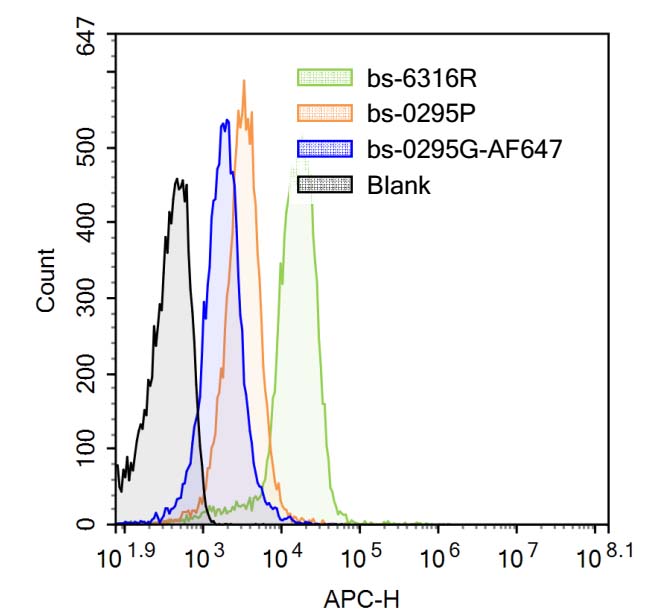 Blank control (Black line): Mouse blood (Black).
Primary Antibody (green line): Rabbit Anti-Prostaglandin E Receptor EP1 antibody (SL6316R)
Dilution: 3μg /10^6 cells;
Isotype Control Antibody (orange line): Rabbit IgG .
Secondary Antibody (white blue line): Goat anti-rabbit IgG-AF647
Dilution: 1μg /test.
Protocol
The cells were fixed with 4% PFA (10min at room temperature)and then were incubated in 5%BSA to block non-specific protein-protein interactions for 30 min at room temperature .Cells stained with Primary Antibody for 30 min at room temperature. The secondary antibody used for 40 min at room temperature. Acquisition of 20,000 events was performed.
Cartpieces
Totalgoods,subtotals:¥Checkout
References (0)
No References
Bought notes(bought amounts latest0)
No one bought this product
User Comment(Total0User Comment Num)
- No comment


 +86 571 56623320
 +86 18668110335

