
Rabbit Anti-TRAK1 antibody
106 kDa O-GlcNAc transferase-interacting protein; 106 kDa O GlcNAc transferase interacting protein; KIAA1042; OGT(O Glc NAc transferase) interacting protein 106 KDa; OIP106; TRAK1_HUMAN; Trafficking kinesin binding protein 1; Trafficking kinesin-binding p
View History [Clear]
Details
Product Name TRAK1 Chinese Name 驱动蛋白Binding protein1抗体 Alias 106 kDa O-GlcNAc transferase-interacting protein; 106 kDa O GlcNAc transferase interacting protein; KIAA1042; OGT(O Glc NAc transferase) interacting protein 106 KDa; OIP106; TRAK1_HUMAN; Trafficking kinesin binding protein 1; Trafficking kinesin-binding protein 1; Trafficking protein kinesin binding 1; TRAK 1. Research Area Tumour immunology Signal transduction Cytoskeleton TumourCell biologyMaker Mitochondrion Immunogen Species Rabbit Clonality Polyclonal React Species Human, Mouse, (predicted: Rat, Chicken, Dog, Pig, Cow, Horse, ) Applications ELISA=1:5000-10000 IHC-P=1:100-500 IHC-F=1:100-500 Flow-Cyt=1ug/Test ICC=1:100 IF=1:100-500 (Paraffin sections need antigen repair)
not yet tested in other applications.
optimal dilutions/concentrations should be determined by the end user.Theoretical molecular weight 106kDa Cellular localization The nucleus cytoplasmic Mitochondrion Form Liquid Concentration 1mg/ml immunogen KLH conjugated synthetic peptide derived from human TRAK1: 171-270/953 Lsotype IgG Purification affinity purified by Protein A Buffer Solution 0.01M TBS(pH7.4) with 1% BSA, 0.03% Proclin300 and 50% Glycerol. Storage Shipped at 4℃. Store at -20 °C for one year. Avoid repeated freeze/thaw cycles. Attention This product as supplied is intended for research use only, not for use in human, therapeutic or diagnostic applications. PubMed PubMed Product Detail The specific function of TRAK1 remains unknown. It interacts with O-GlcNAc transferase, RHOT1/Miro-1 and RHOT2/Miro-2. It shows high expression in spinal cord and moderate expression in all other tissues and specific brain regions examined.
Function:
Involved in the regulation of endosome-to-lysosome trafficking, including endocytic trafficking of EGF-EGFR complexes and GABA-A receptors.
Subunit:
Interacts with O-GlcNAc transferase. Interacts with RHOT1/Miro-1 and RHOT2/Miro-2. Interacts with HGS. Interacts with GABRA1. Interacts with KIF5C.
Subcellular Location:
Cytoplasm. Nucleus. Mitochondrion. Early endosome. Endosome.
Tissue Specificity:
High expression in spinal cord and moderate expression in all other tissues and specific brain regions examined. Expressed in all cell lines examined.
Post-translational modifications:
O-glycosylated.
Similarity:
Contains 1 HAP1 N-terminal domain.
SWISS:
Q9UPV9
Gene ID:
22906
Database links:Entrez Gene: 22906 Human
Omim: 608112 Human
SwissProt: Q9UPV9 Human
Unigene: 535711 Human
Product Picture 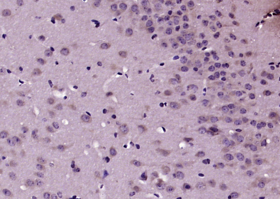 Paraformaldehyde-fixed, paraffin embedded (mouse brain tissue); Antigen retrieval by boiling in sodium citrate buffer (pH6.0) for 15min; Block endogenous peroxidase by 3% hydrogen peroxide for 20 minutes; Blocking buffer (normal goat serum) at 37°C for 30min; Antibody incubation with (TRAK1) Polyclonal Antibody, Unconjugated (SL5536R) at 1:200 overnight at 4°C, followed by operating according to SP Kit(Rabbit) (sp-0023) instructionsand DAB staining.
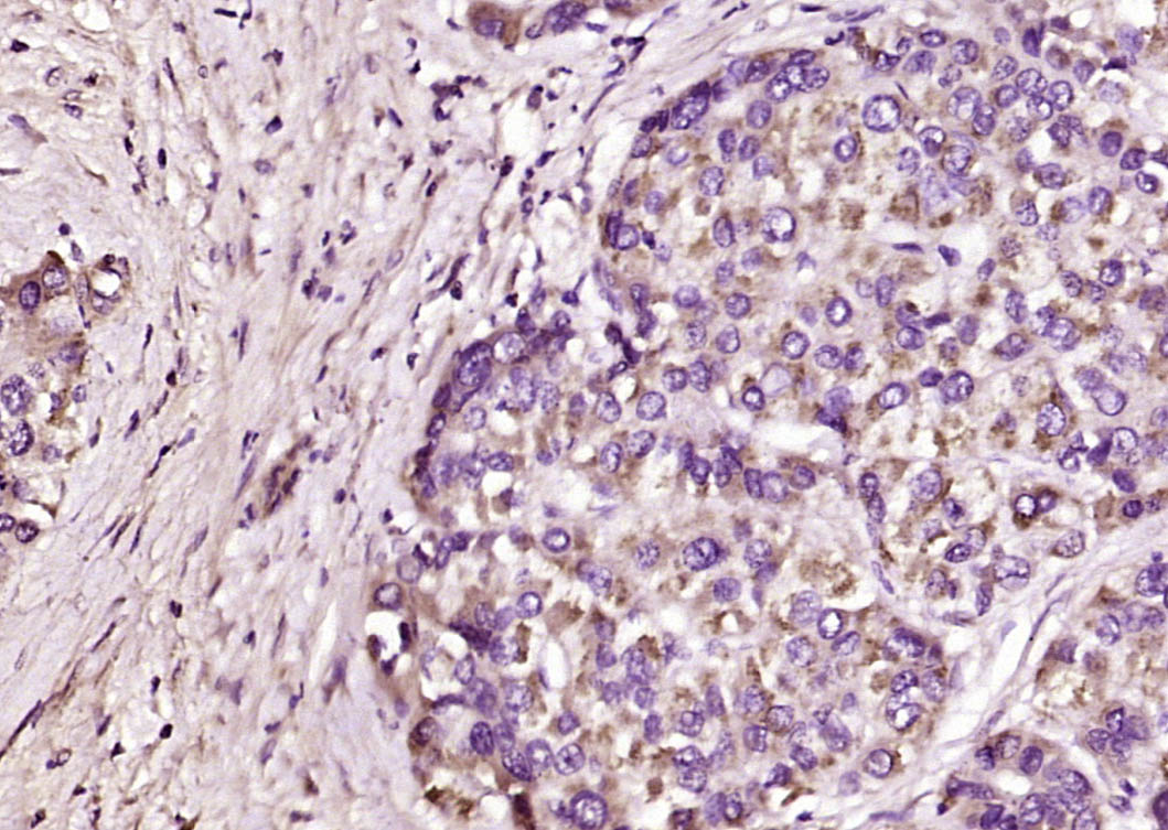 Paraformaldehyde-fixed, paraffin embedded (human liver cancer); Antigen retrieval by boiling in sodium citrate buffer (pH6.0) for 15min; Block endogenous peroxidase by 3% hydrogen peroxide for 20 minutes; Blocking buffer (normal goat serum) at 37°C for 30min; Antibody incubation with (TRAK1) Polyclonal Antibody, Unconjugated (SL5536R) at 1:200 overnight at 4°C, followed by operating according to SP Kit(Rabbit) (sp-0023) instructionsand DAB staining.
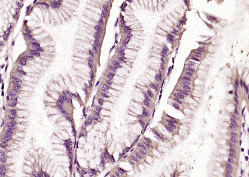 Paraformaldehyde-fixed, paraffin embedded (human gastric carcinoma); Antigen retrieval by boiling in sodium citrate buffer (pH6.0) for 15min; Block endogenous peroxidase by 3% hydrogen peroxide for 20 minutes; Blocking buffer (normal goat serum) at 37°C for 30min; Antibody incubation with (TRAK1) Polyclonal Antibody, Unconjugated (SL5536R) at 1:200 overnight at 4°C, followed by operating according to SP Kit(Rabbit) (sp-0023) instructionsand DAB staining.
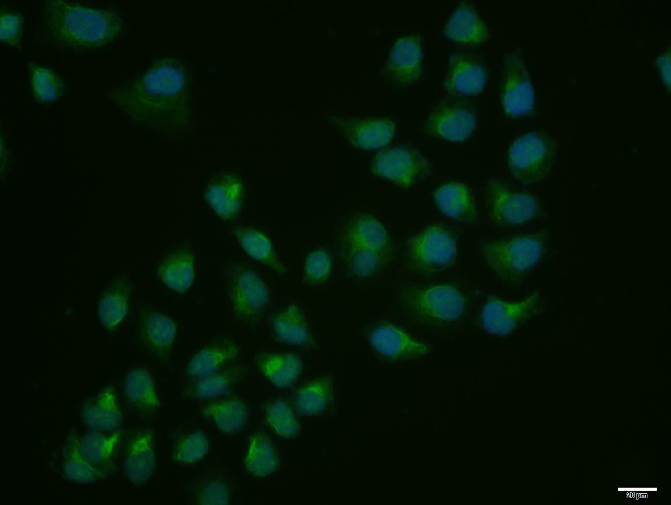 Hela cell; 4% Paraformaldehyde-fixed; Triton X-100 at room temperature for 20 min; Blocking buffer (normal goat serum, C-0005) at 37°C for 20 min; Antibody incubation with (TRAK1) polyclonal Antibody, Unconjugated (SL5536R) 1:100, 90 minutes at 37°C; followed by a conjugated Goat Anti-Rabbit IgG antibody at 37°C for 90 minutes, DAPI (blue, C02-04002) was used to stain the cell nuclei.
Cartpieces
Totalgoods,subtotals:¥Checkout
Bought notes(bought amounts latest0)
No one bought this product
User Comment(Total0User Comment Num)
- No comment


 +86 571 56623320
 +86 18668110335

