
Rabbit Anti-phospho-ROCK1 (Thr455+Ser456)antibody
p-ROCK1(Thr455/Ser456); p160 ROCK1; p160ROCK; Renal carcinoma antigen NY REN 35; Rho associated coiled coil containing protein kinase 1; Rho associated protein kinase 1; Rho-associated coiled-coil containing protein kinase 1; ROCK1_HUMAN.
View History [Clear]
Details
Product Name phospho-ROCK1 (Thr455+Ser456) Chinese Name 磷酸化Rho相关蛋白激酶1抗体 Alias p-ROCK1(Thr455/Ser456); p160 ROCK1; p160ROCK; Renal carcinoma antigen NY REN 35; Rho associated coiled coil containing protein kinase 1; Rho associated protein kinase 1; Rho-associated coiled-coil containing protein kinase 1; ROCK1_HUMAN. literatures Product Type Phosphorylated anti Research Area Tumour Signal transduction Kinases and Phosphatases Immunogen Species Rabbit Clonality Polyclonal React Species Human, Mouse, (predicted: Rat, Dog, Pig, Horse, Rabbit, ) Applications WB=1:500-2000 ELISA=1:5000-10000 IHC-P=1:100-500 IHC-F=1:100-500 Flow-Cyt=1ug/Test IF=1:100-500 (Paraffin sections need antigen repair)
not yet tested in other applications.
optimal dilutions/concentrations should be determined by the end user.Theoretical molecular weight 158kDa Cellular localization cytoplasmic The cell membrane Form Liquid Concentration 1mg/ml immunogen KLH conjugated synthesised phosphopeptide derived from human ROCK1 around the phosphorylation site of Thr455+Ser456: CR(p-T)(p-S)N Lsotype IgG Purification affinity purified by Protein A Buffer Solution 0.01M TBS(pH7.4) with 1% BSA, 0.03% Proclin300 and 50% Glycerol. Storage Shipped at 4℃. Store at -20 °C for one year. Avoid repeated freeze/thaw cycles. Attention This product as supplied is intended for research use only, not for use in human, therapeutic or diagnostic applications. PubMed PubMed Product Detail This gene encodes a protein serine/threonine kinase that is activated when bound to the GTP-bound form of Rho. The small GTPase Rho regulates formation of focal adhesions and stress fibers of fibroblasts, as well as adhesion and aggregation of platelets and lymphocytes by shuttling between the inactive GDP-bound form and the active GTP-bound form. Rho is also essential in cytokinesis and plays a role in transcriptional activation by serum response factor. This protein, a downstream effector of Rho, phosphorylates and activates LIM kinase, which in turn, phosphorylates cofilin, inhibiting its actin-depolymerizing activity. [provided by RefSeq].
Function:
Protein kinase which is a key regulator of actin cytoskeleton and cell polarity. Involved in regulation of smooth muscle contraction, actin cytoskeleton organization, stress fiber and focal adhesion formation, neurite retraction, cell adhesion and motility via phosphorylation of DAPK3, GFAP, LIMK1, LIMK2, MYL9/MLC2, PFN1 and PPP1R12A. Phosphorylates FHOD1 and acts synergistically with it to promote SRC-dependent non-apoptotic plasma membrane blebbing. Phosphorylates JIP3 and regulates the recruitment of JNK to JIP3 upon UVB-induced stress. Acts as a suppressor of inflammatory cell migration by regulating PTEN phosphorylation and stability. Acts as a negative regulator of VEGF-induced angiogenic endothelial cell activation. Required for centrosome positioning and centrosome-dependent exit from mitosis. Plays a role in terminal erythroid differentiation. May regulate closure of the eyelids and ventral body wall by inducing the assembly of actomyosin bundles. Promotes keratinocyte terminal differentiation.
Subunit:
Homodimer. Interacts with RHOB, RHOC, MYLC2B snd PTEN (By similarity). Interacts with RHOA (activated by GTP), CHORDC1, DAPK3, GEM, JIP3, RHOE, PPP1R12A, PFN1, LIMK1, LIMK2 and TSG101. Interacts with FHOD1 in a Src-dependent manner.
Subcellular Location:
Cytoplasm. Cytoplasm, cytoskeleton, centrosome, centriole (By similarity). Golgi apparatus membrane; Peripheral membrane protein. Cell projection, bleb. Note=Associated with the mother centriole and an intercentriolar linker (By similarity). A small proportion is associated with Golgi membranes.
Tissue Specificity:
Detected in blood platelets.
Post-translational modifications:
Autophosphorylated on serine and threonine residues. Phosphorylated upon DNA damage, probably by ATM or ATR.
Cleaved by caspase-3 during apoptosis. This leads to constitutive activation of the kinase and membrane blebbing.
Similarity:
Belongs to the protein kinase superfamily. AGC Ser/Thr protein kinase family.
Contains 1 AGC-kinase C-terminal domain.
Contains 1 PH domain.
Contains 1 phorbol-ester/DAG-type zinc finger.
Contains 1 protein kinase domain.
Contains 1 REM (Hr1) repeat.
SWISS:
Q13464
Gene ID:
6093
Database links:Entrez Gene: 6093 Human
Entrez Gene: 19877 Mouse
Omim: 601702 Human
SwissProt: Q13464 Human
SwissProt: P70335 Mouse
ROCK1也是丝/苏氨基酸激酶的一种,可调节cell factor,促进平滑肌收缩和肌动蛋白的形成,因此又称:含有卷曲螺旋的蛋白激酶。目前主要用于Tumour浸润、转移方面的研究。Product Picture 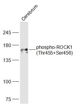 Sample:
Cerebrum (Mouse) Lysate at 40 ug
Primary: Anti-phospho-ROCK1(Thr455+Ser456) (SL4630R) at 1/1000 dilution
Secondary: IRDye800CW Goat Anti-Rabbit IgG at 1/20000 dilution
Predicted band size: 158 kD
Observed band size: 158 kD
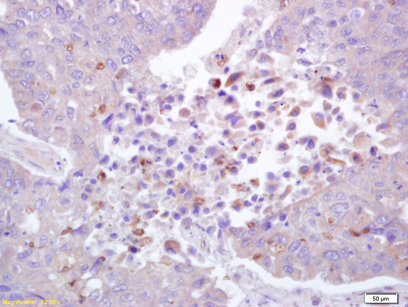 Tissue/cell: human lung carcinoma; 4% Paraformaldehyde-fixed and paraffin-embedded;
Antigen retrieval: citrate buffer ( 0.01M, pH 6.0 ), Boiling bathing for 15min; Block endogenous peroxidase by 3% Hydrogen peroxide for 30min; Blocking buffer (normal goat serum,C-0005) at 37℃ for 20 min;
Incubation: Anti-phospho-ROCK1(Thr455/Ser456) Polyclonal Antibody, Unconjugated(SL4630R) 1:200, overnight at 4°C, followed by conjugation to the secondary antibody(SP-0023) and DAB(C-0010) staining
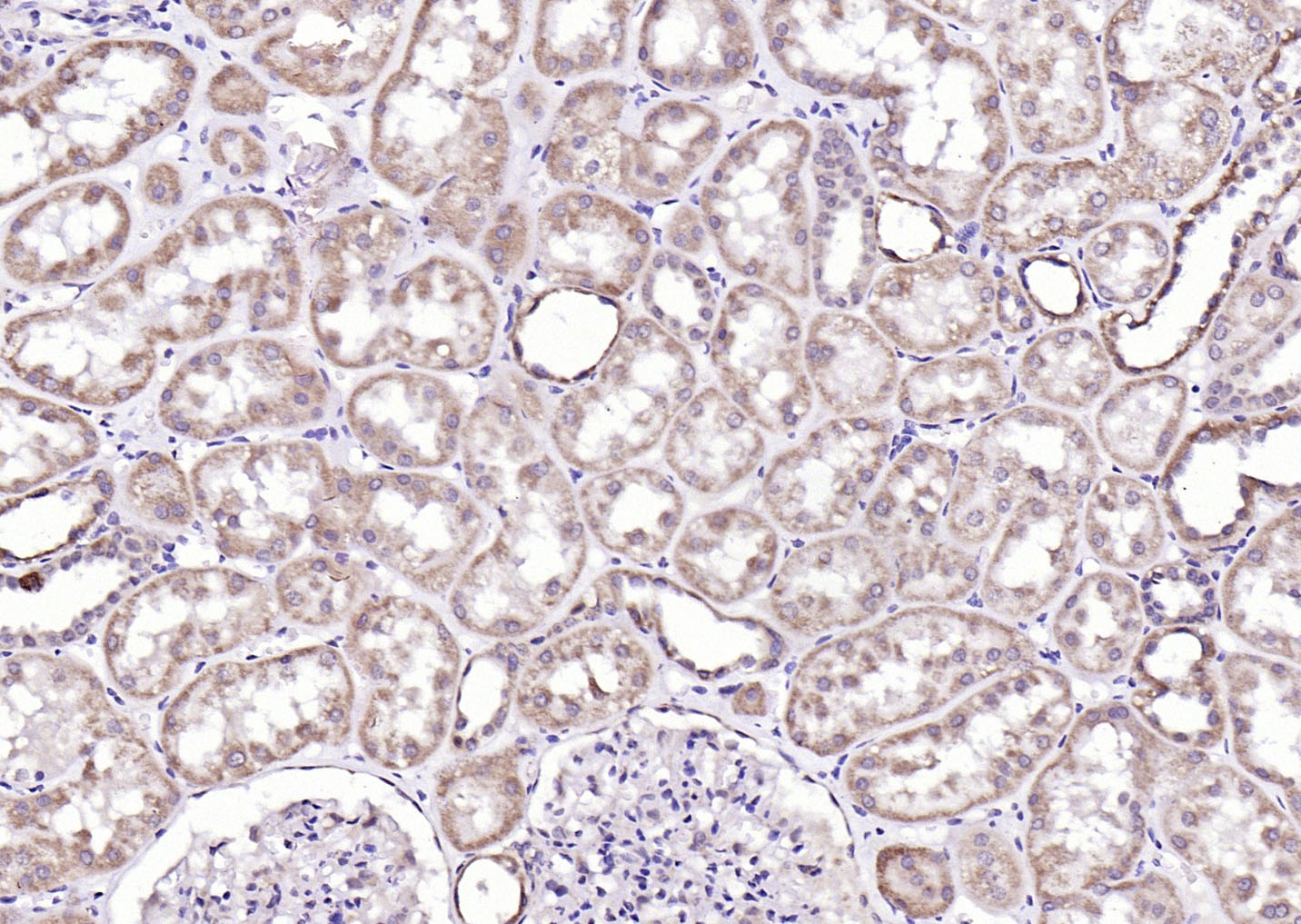 Paraformaldehyde-fixed, paraffin embedded (Human kidney); Antigen retrieval by boiling in sodium citrate buffer (pH6.0) for 15min; Block endogenous peroxidase by 3% hydrogen peroxide for 20 minutes; Blocking buffer (normal goat serum) at 37°C for 30min; Antibody incubation with (phospho-ROCK1 (Thr455+Ser456)) Polyclonal Antibody, Unconjugated (SL4630R) at 1:200 overnight at 4°C, followed by operating according to SP Kit(Rabbit) (sp-0023) instructionsand DAB staining.
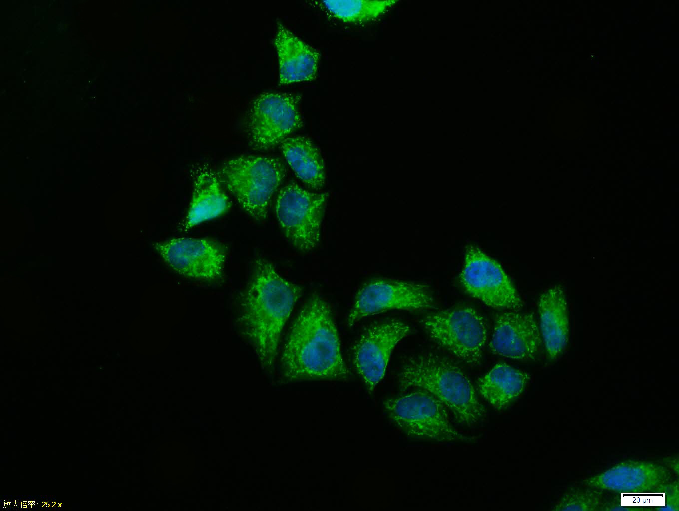 Hela cell; 4% Paraformaldehyde-fixed; Triton X-100 at room temperature for 20 min; Blocking buffer (normal goat serum, C-0005) at 37°C for 20 min; Antibody incubation with (phospho-ROCK1 (Thr455+Ser456)) polyclonal Antibody, Unconjugated (SL4630R) 1:100, 90 minutes at 37°C; followed by a conjugated Goat Anti-Rabbit IgG antibody at 37°C for 90 minutes, DAPI (blue, C02-04002) was used to stain the cell nuclei.
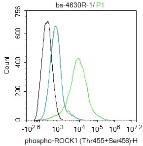 Blank control:MCF-7.
Primary Antibody (green line): Rabbit Anti-phospho-ROCK1 (Thr455+Ser456) antibody (SL4630R)
Dilution: 1ug/Test;
Secondary Antibody (white blue line) : Goat anti-rabbit IgG-AF488
Dilution: 0.5ug/Test.
Isotype control(orange line):Normal Rabbit IgG
Protocol
The cells were incubated in 5%BSA to block non-specific protein-protein interactions for 30 min at room temperature .Cells stained with Primary Antibody for 30 min at room temperature. The secondary antibody used for 40 min at room temperature. Acquisition of 20,000 events was performed. Blank control:MCF-7.
Primary Antibody (green line): Rabbit Anti-phospho-ROCK1 (Thr455+Ser456) antibody (SL4630R)
Dilution: 1ug/Test;
Secondary Antibody (white blue line) : Goat anti-rabbit IgG-AF488
Dilution: 0.5ug/Test.
Isotype control(orange line):Normal Rabbit IgG
Protocol
The cells were incubated in 5%BSA to block non-specific protein-protein interactions for 30 min at room temperature .Cells stained with Primary Antibody for 30 min at room temperature. The secondary antibody used for 40 min at room temperature. Acquisition of 20,000 events was performed.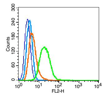 Blank control(blue): Hela (fixed with 2% paraformaldehyde (10 min) , then permeabilized with 90% ice-cold methanol for 30 min on ice).
Primary Antibody:Rabbit Anti- phospho-ROCK1(Thr455+Ser456) antibody(SL4630R), Dilution:1μg in 100 μL 1X PBS containing 0.5% BSA;
Isotype Control Antibody: Rabbit IgG(orange) ,used under the same conditions );
Secondary Antibody: Goat anti-rabbit IgG-PE(white blue), Dilution: 1:200 in 1 X PBS containing 0.5% BSA.
Cartpieces
Totalgoods,subtotals:¥Checkout
References (0)
No References
Bought notes(bought amounts latest0)
No one bought this product
User Comment(Total0User Comment Num)
- No comment


 +86 571 56623320
 +86 18668110335

