
Rabbit Anti-SGPL1 antibody
FLJ13811; hSPL; KIAA1252; S1P; S1PL; SGPL 1; SGPL1; SGPL1_HUMAN; SP lyase; SP-lyase 1; Sphingosine 1 phosphate aldolase; Sphingosine 1 phosphate lyase 1; Sphingosine-1-phosphate aldolase; Sphingosine-1-phosphate lyase 1; SPL 1; SPL.
View History [Clear]
Details
Product Name SGPL1 Chinese Name 磷酸鞘氨醇裂解酶1抗体 Alias FLJ13811; hSPL; KIAA1252; S1P; S1PL; SGPL 1; SGPL1; SGPL1_HUMAN; SP lyase; SP-lyase 1; Sphingosine 1 phosphate aldolase; Sphingosine 1 phosphate lyase 1; Sphingosine-1-phosphate aldolase; Sphingosine-1-phosphate lyase 1; SPL 1; SPL. literatures Research Area Tumour Cardiovascular immunology Signal transduction Lipoprotein The new supersedes the old Immunogen Species Rabbit Clonality Polyclonal React Species Human, Mouse, Rat, (predicted: Dog, Pig, Cow, Horse, Rabbit, ) Applications ELISA=1:5000-10000 IHC-P=1:100-500 IHC-F=1:100-500 Flow-Cyt=1ug/test IF=1:100-500 (Paraffin sections need antigen repair)
not yet tested in other applications.
optimal dilutions/concentrations should be determined by the end user.Theoretical molecular weight 63kDa Cellular localization cytoplasmic Form Liquid Concentration 1mg/ml immunogen KLH conjugated synthetic peptide derived from human SGPL1: 301-400/568 Lsotype IgG Purification affinity purified by Protein A Buffer Solution 0.01M TBS(pH7.4) with 1% BSA, 0.03% Proclin300 and 50% Glycerol. Storage Shipped at 4℃. Store at -20 °C for one year. Avoid repeated freeze/thaw cycles. Attention This product as supplied is intended for research use only, not for use in human, therapeutic or diagnostic applications. PubMed PubMed Product Detail Cleaves phosphorylated sphingoid bases (PSBs), such as sphingosine-1-phosphate, into fatty aldehydes and phosphoethanolamine. Elevates stress-induced ceramide production and apoptosis.
Function:
Cleaves phosphorylated sphingoid bases (PSBs), such as sphingosine-1-phosphate, into fatty aldehydes and phosphoethanolamine. Elevates stress-induced ceramide production and apoptosis.
Subcellular Location:
Endoplasmic reticulum membrane; Single-pass type III membrane protein.
Tissue Specificity:
Found in liver and kidney.
Similarity:
Belongs to the group II decarboxylase family. Sphingosine-1-phosphate lyase subfamily.
SWISS:
O95470
Gene ID:
8879
Database links:Entrez Gene: 8879 Human
Entrez Gene: 20397 Mouse
SwissProt: O95470 Human
SwissProt: Q8R0X7 Mouse
Unigene: 499984 Human
Unigene: 412319 Mouse
Unigene: 431862 Mouse
Unigene: 26953 Rat
Omim: 603729 Human
Product Picture 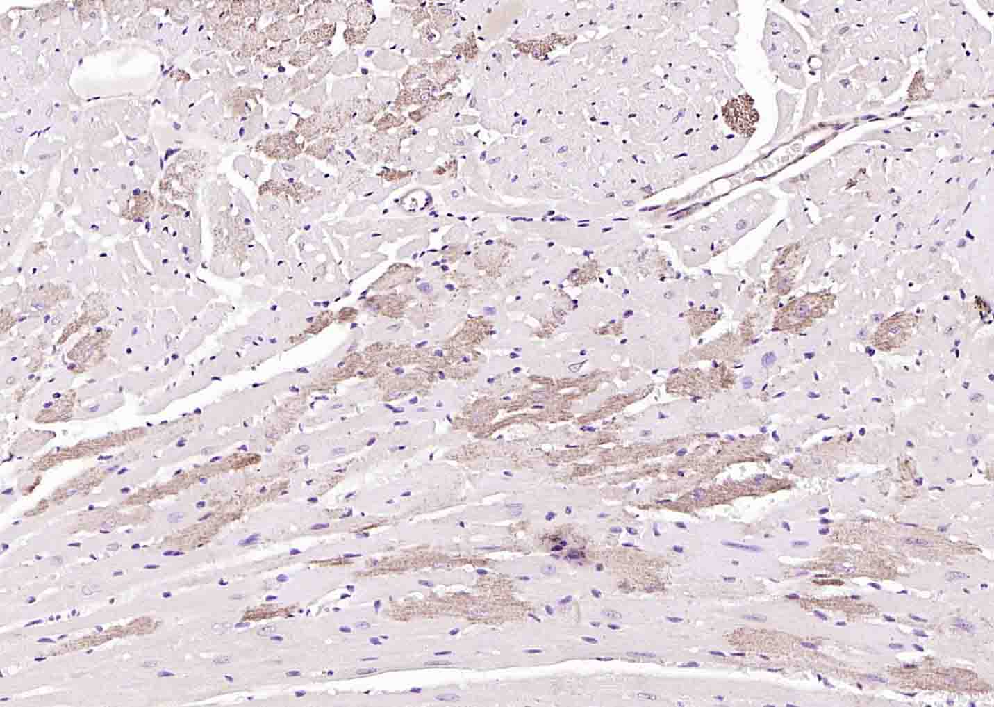 Paraformaldehyde-fixed, paraffin embedded (mouse heart); Antigen retrieval by boiling in sodium citrate buffer (pH6.0) for 15min; Block endogenous peroxidase by 3% hydrogen peroxide for 20 minutes; Blocking buffer (normal goat serum) at 37°C for 30min; Antibody incubation with (SGPL1) Polyclonal Antibody, Unconjugated (SL4188R) at 1:200 overnight at 4°C, followed by operating according to SP Kit(Rabbit) (sp-0023) instructionsand DAB staining.
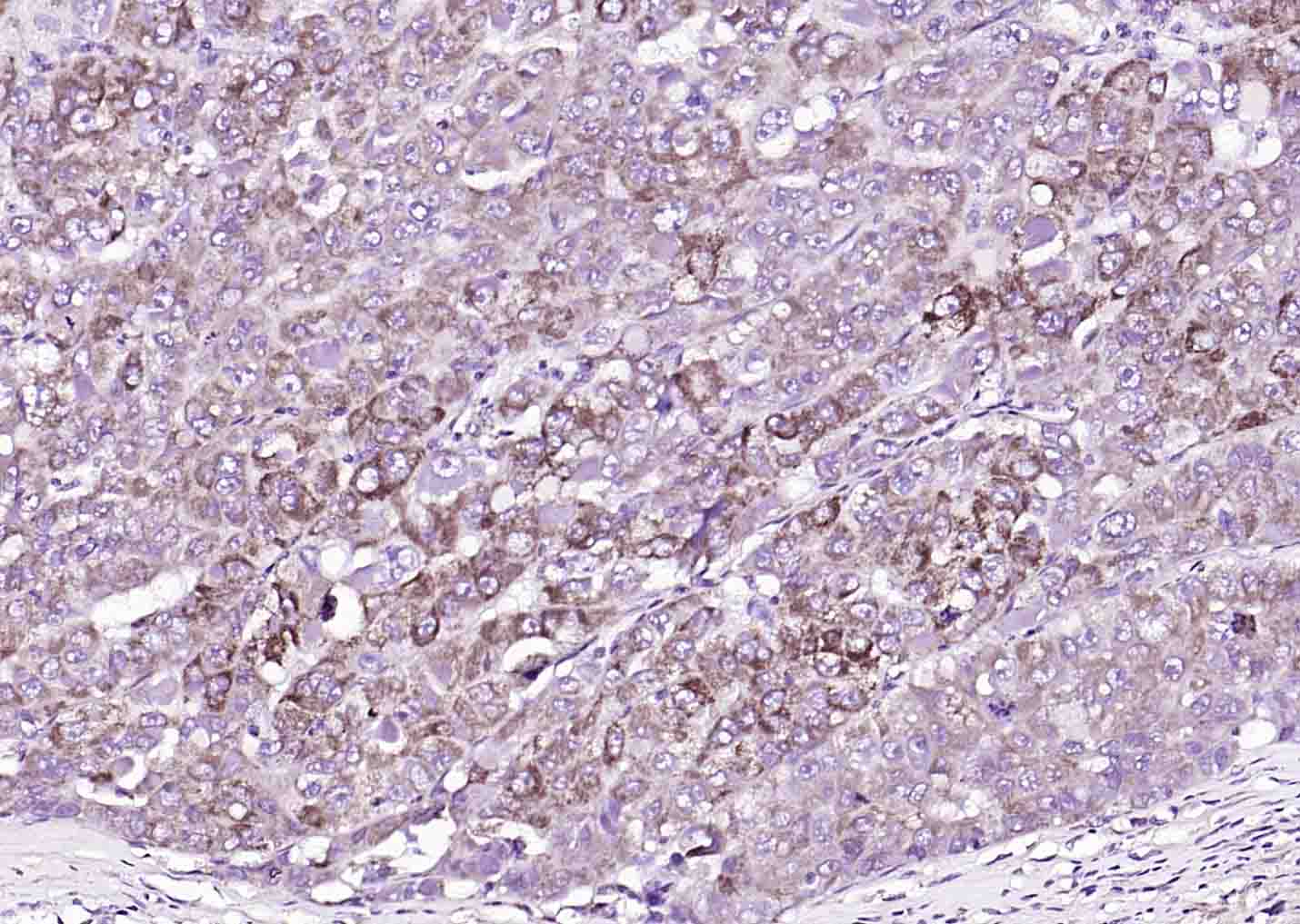 Paraformaldehyde-fixed, paraffin embedded (human liver carcinoma); Antigen retrieval by boiling in sodium citrate buffer (pH6.0) for 15min; Block endogenous peroxidase by 3% hydrogen peroxide for 20 minutes; Blocking buffer (normal goat serum) at 37°C for 30min; Antibody incubation with (SGPL1) Polyclonal Antibody, Unconjugated (SL4188R) at 1:200 overnight at 4°C, followed by operating according to SP Kit(Rabbit) (sp-0023) instructionsand DAB staining.
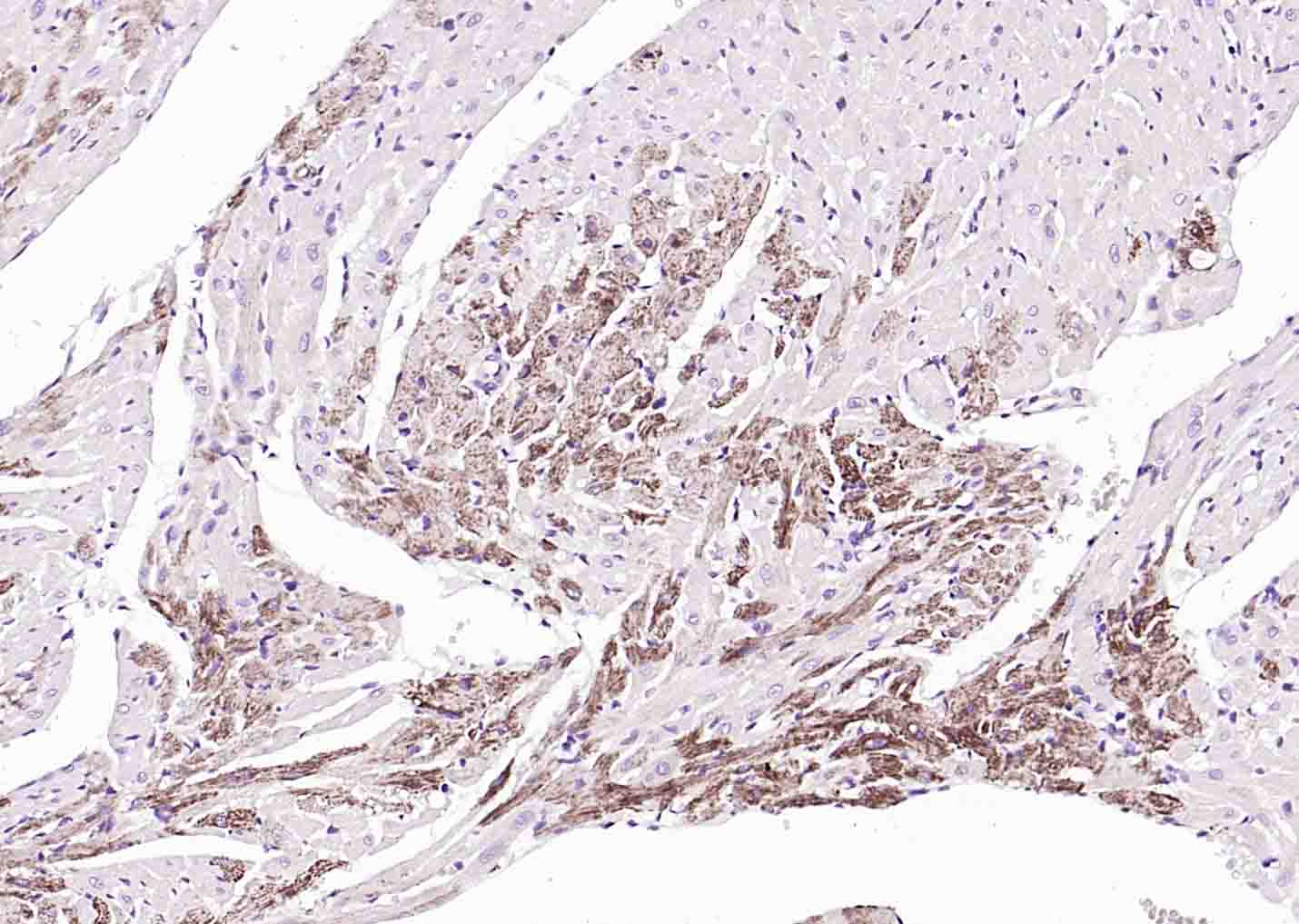 Paraformaldehyde-fixed, paraffin embedded (rat heart); Antigen retrieval by boiling in sodium citrate buffer (pH6.0) for 15min; Block endogenous peroxidase by 3% hydrogen peroxide for 20 minutes; Blocking buffer (normal goat serum) at 37°C for 30min; Antibody incubation with (SGPL1) Polyclonal Antibody, Unconjugated (SL4188R) at 1:200 overnight at 4°C, followed by operating according to SP Kit(Rabbit) (sp-0023) instructionsand DAB staining.
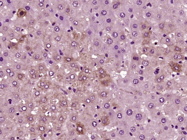 Paraformaldehyde-fixed, paraffin embedded (Rat liver); Antigen retrieval by boiling in sodium citrate buffer (pH6.0) for 15min; Block endogenous peroxidase by 3% hydrogen peroxide for 20 minutes; Blocking buffer (normal goat serum) at 37°C for 30min; Antibody incubation with (SGPL1) Polyclonal Antibody, Unconjugated (SL4188R) at 1:400 overnight at 4°C, followed by operating according to SP Kit(Rabbit) (sp-0023) instructions and DAB staining.
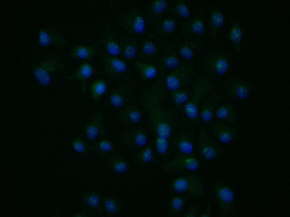 HepG2 cell; 4% Paraformaldehyde-fixed; Triton X-100 at room temperature for 20 min; Blocking buffer (normal goat serum, C-0005) at 37°C for 20 min; Antibody incubation with (SGPL1) polyclonal Antibody, Unconjugated (SL4188R) 1:100, 90 minutes at 37°C; followed by a conjugated Goat Anti-Rabbit IgG antibody at 37°C for 90 minutes, DAPI (blue, C02-04002) was used to stain the cell nuclei.
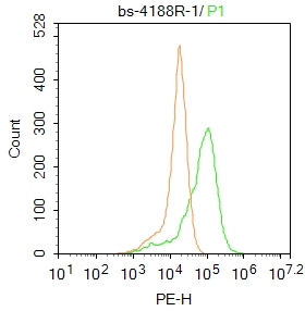 Blank control: A549.
Primary Antibody (green line): Rabbit Anti-SGPL1 antibody (SL4188R)
Dilution: 1μg /10^6 cells;
Isotype Control Antibody (orange line): Rabbit IgG .
Secondary Antibody : Goat anti-rabbit IgG-PE
Dilution: 1μg /test.
Protocol
The cells were fixed with 4% PFA (10min at room temperature)and then permeabilized with PBST for 20 min at room temperature. The cells were then incubated in 5%BSA to block non-specific protein-protein interactions for 30 min at room temperature .Cells stained with Primary Antibody for 30 min at room temperature. The secondary antibody used for 40 min at room temperature. Acquisition of 20,000 events was performed.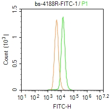 Blank control: A549.
Primary Antibody (green line): Rabbit Anti-SGPL1/FITC Conjugated antibody (SL4188R-FITC)
Dilution: 1μg /10^6 cells;
Isotype Control Antibody (orange line): Rabbit IgG-FITC .
Protocol
The cells were fixed with 4% PFA (10min at room temperature)and then permeabilized with 0.1% PBST for 20 min at-20℃. The cells were then incubated in 5% BSA to block non-specific protein-protein interactions for 30 min at room temperature. The cells were stained with Primary Antibody for 30 min at room temperature. Acquisition of 20,000 events was performed.
Cartpieces
Totalgoods,subtotals:¥Checkout
References (0)
No References
Bought notes(bought amounts latest0)
No one bought this product
User Comment(Total0User Comment Num)
- No comment


 +86 571 56623320
 +86 18668110335

