
Rabbit Anti-PI 3 Kinase Class 3 antibody
PI 3 Kinase Class 3; Phosphatidylinositol 3 kinase catalytic subunit type 3; Phosphatidylinositol 3 kinase class 3; Phosphatidylinositol 3 kinase p100 subunit; Phosphoinositide 3 kinase class 3; PI3 kinase type 3; PI3K type 3; PIK3C3; PtdIns 3 kinase type
View History [Clear]
Details
Product Name PI 3 Kinase Class 3 Chinese Name 磷脂酰肌醇激酶3催化亚单位3抗体 Alias PI 3 Kinase Class 3; Phosphatidylinositol 3 kinase catalytic subunit type 3; Phosphatidylinositol 3 kinase class 3; Phosphatidylinositol 3 kinase p100 subunit; Phosphoinositide 3 kinase class 3; PI3 kinase type 3; PI3K type 3; PIK3C3; PtdIns 3 kinase type 3; Vps 34; Vps34; hVps34; MGC61518; PIK3C3; PI3 Kinase p100; PK3C3_HUMAN. Research Area immunology Signal transduction Kinases and Phosphatases Immunogen Species Rabbit Clonality Polyclonal React Species Human, Mouse, Rat, (predicted: Chicken, Dog, Pig, Cow, Horse, Rabbit, ) Applications WB=1:500-2000 ELISA=1:5000-10000 IHC-P=1:100-500 IHC-F=1:100-500 IF=1:100-500 (Paraffin sections need antigen repair)
not yet tested in other applications.
optimal dilutions/concentrations should be determined by the end user.Theoretical molecular weight 98kDa Cellular localization cytoplasmic Form Liquid Concentration 1mg/ml immunogen KLH conjugated synthetic peptide derived from human PIK3C3/PI3 kinase type 3: 301-400/887 Lsotype IgG Purification affinity purified by Protein A Buffer Solution 0.01M TBS(pH7.4) with 1% BSA, 0.03% Proclin300 and 50% Glycerol. Storage Shipped at 4℃. Store at -20 °C for one year. Avoid repeated freeze/thaw cycles. Attention This product as supplied is intended for research use only, not for use in human, therapeutic or diagnostic applications. PubMed PubMed Product Detail PI 3 Kinase Class 3 is a member of the PI3/PI4-kinase family. It is the catalytic subunit of the PI3K complex. It is involved in the transport of lysosomal enzyme precursors to lysosomes. It is ubiquitously expressed, with a highest expression in skeletal muscle.
Function:
Catalytic subunit of the PI3K complex that mediates formation of phosphatidylinositol 3-phosphate which plays a key role in initiation and maturation of autophagosomes. Involved in the transport of lysosomal enzyme precursors to lysosomes. Required for the abcission step in cytokinesis. Required for transport from early to late endosomes.
Subunit:
Heterodimer. This subunit, part of a complex composed of regulatory and catalytic subunits, associates with regulatory subunit PIK3R4. Forms a complex with BECN1, PIK3R4 and either UVRAG and KIAA0226/Rubicon, or with ATG14. In this complex, presence of UVRAG and ATG14 are mutually exclusive. Part of a complex composed of PIK3R4 and PIK3CB (By similarity). Interacts with RAB7A in the presence of PIK3R4.
Subcellular Location:
Midbody. Late endosome.
Tissue Specificity:
Ubiquitously expressed, with a highest expression in skeletal muscle.
Similarity:
Belongs to the PI3/PI4-kinase family.
Contains 1 C2 PI3K-type domain.
Contains 1 PI3K/PI4K domain.
Contains 1 PIK helical domain.
SWISS:
Q8NEB9
Gene ID:
5289
Database links:Entrez Gene: 5289 Human
Entrez Gene: 225326 Mouse
Omim: 602609 Human
SwissProt: Q8NEB9 Human
SwissProt: Q6PF93 Mouse
Unigene: 464971 Human
Unigene: 194127 Mouse
Unigene: 30010 Rat
Product Picture 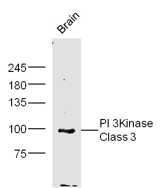 Sample:
brain(mouse) Lysate at 40 ug
Primary: Anti-PI 3 Kinase Class 3 (SL4159R) at 1/300 dilution
Secondary: IRDye800CW Goat Anti-Rabbit IgG at 1/20000 dilution
Predicted band size: 98kD
Observed band size: 98 kD
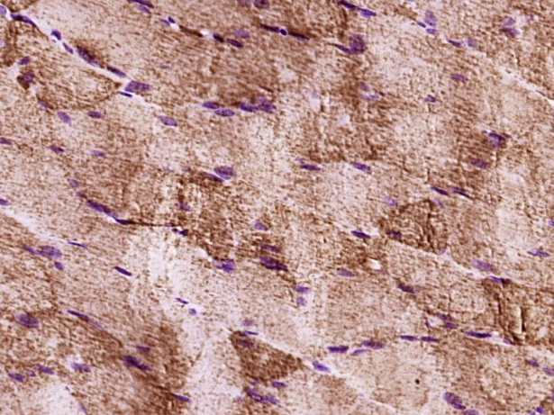 Paraformaldehyde-fixed, paraffin embedded (Rat skeletal muscle); Antigen retrieval by boiling in sodium citrate buffer (pH6.0) for 15min; Block endogenous peroxidase by 3% hydrogen peroxide for 20 minutes; Blocking buffer (normal goat serum) at 37°C for 30min; Antibody incubation with (PI 3 Kinase Class 3) Polyclonal Antibody, Unconjugated (SL4159R) at 1:400 overnight at 4°C, followed by operating according to SP Kit(Rabbit) (sp-0023) instructionsand DAB staining.
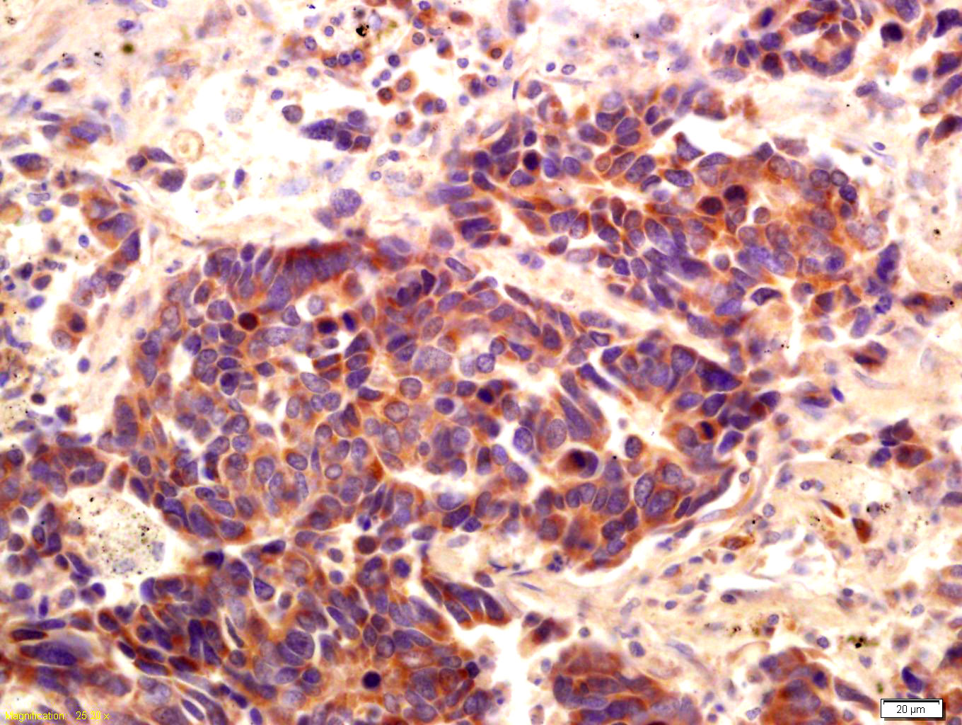 Tissue/cell: human lung carcinoma; 4% Paraformaldehyde-fixed and paraffin-embedded;
Antigen retrieval: citrate buffer ( 0.01M, pH 6.0 ), Boiling bathing for 15min; Block endogenous peroxidase by 3% Hydrogen peroxide for 30min; Blocking buffer (normal goat serum,C-0005) at 37℃ for 20 min;
Incubation: Anti-PI 3 Kinase Class 3 Polyclonal Antibody, Unconjugated(SL4159R) 1:200, overnight at 4°C, followed by conjugation to the secondary antibody(SP-0023) and DAB(C-0010) staining
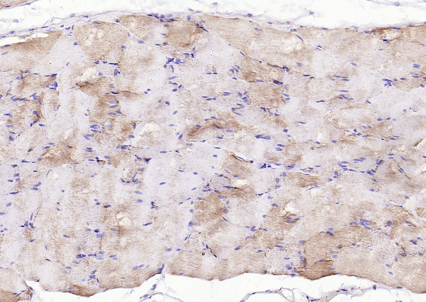 Paraformaldehyde-fixed, paraffin embedded (Rat skeletal muscle); Antigen retrieval by boiling in sodium citrate buffer (pH6.0) for 15min; Block endogenous peroxidase by 3% hydrogen peroxide for 20 minutes; Blocking buffer (normal goat serum) at 37°C for 30min; Antibody incubation with (PI 3 Kinase Class 3) Polyclonal Antibody, Unconjugated (SL4159R) at 1:200 overnight at 4°C, followed by operating according to SP Kit(Rabbit) (sp-0023) instructionsand DAB staining.
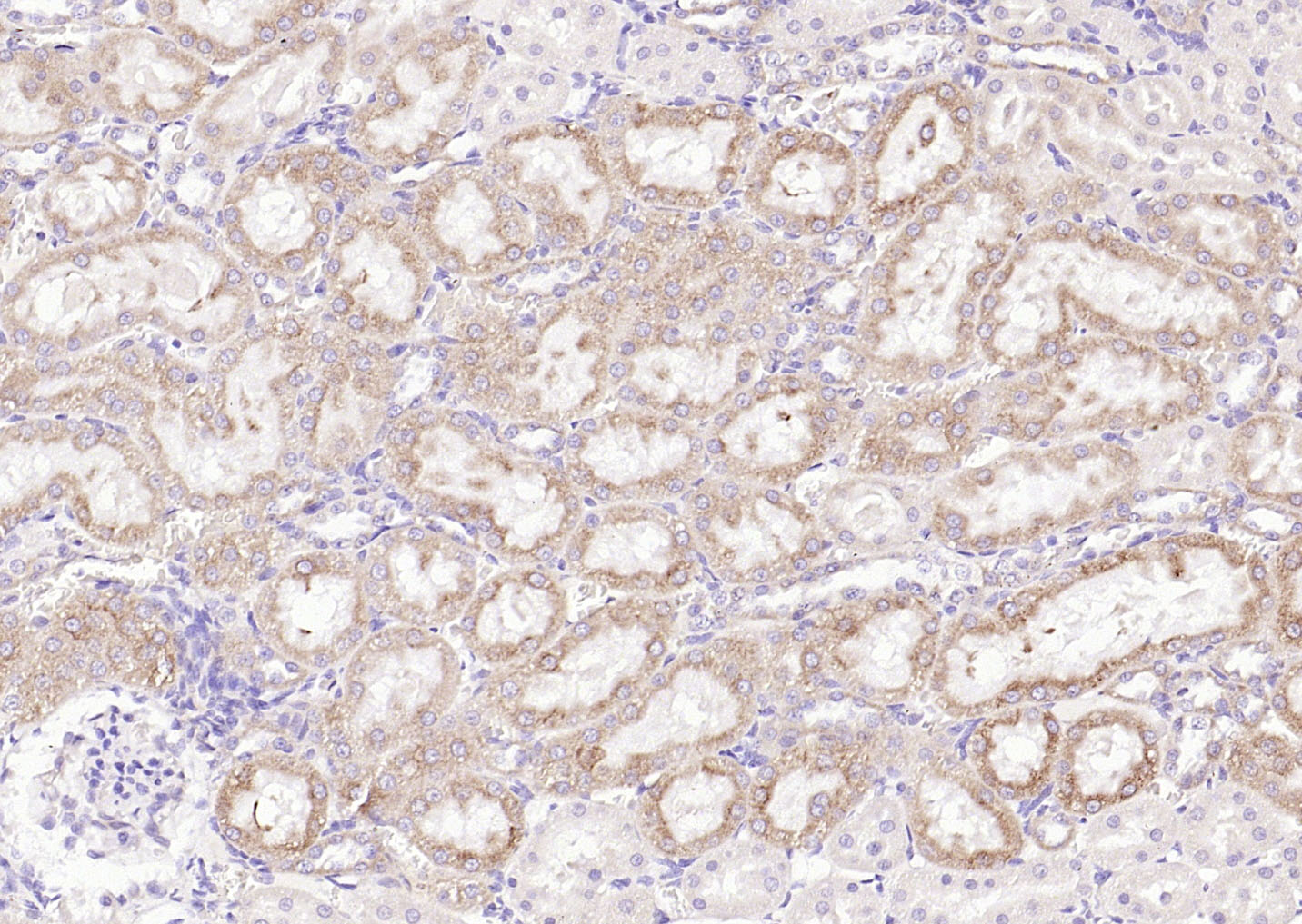 Paraformaldehyde-fixed, paraffin embedded (Rat kidney ); Antigen retrieval by boiling in sodium citrate buffer (pH6.0) for 15min; Block endogenous peroxidase by 3% hydrogen peroxide for 20 minutes; Blocking buffer (normal goat serum) at 37°C for 30min; Antibody incubation with (PI 3 Kinase Class 3) Polyclonal Antibody, Unconjugated (SL4159R) at 1:200 overnight at 4°C, followed by operating according to SP Kit(Rabbit) (sp-0023) instructionsand DAB staining.
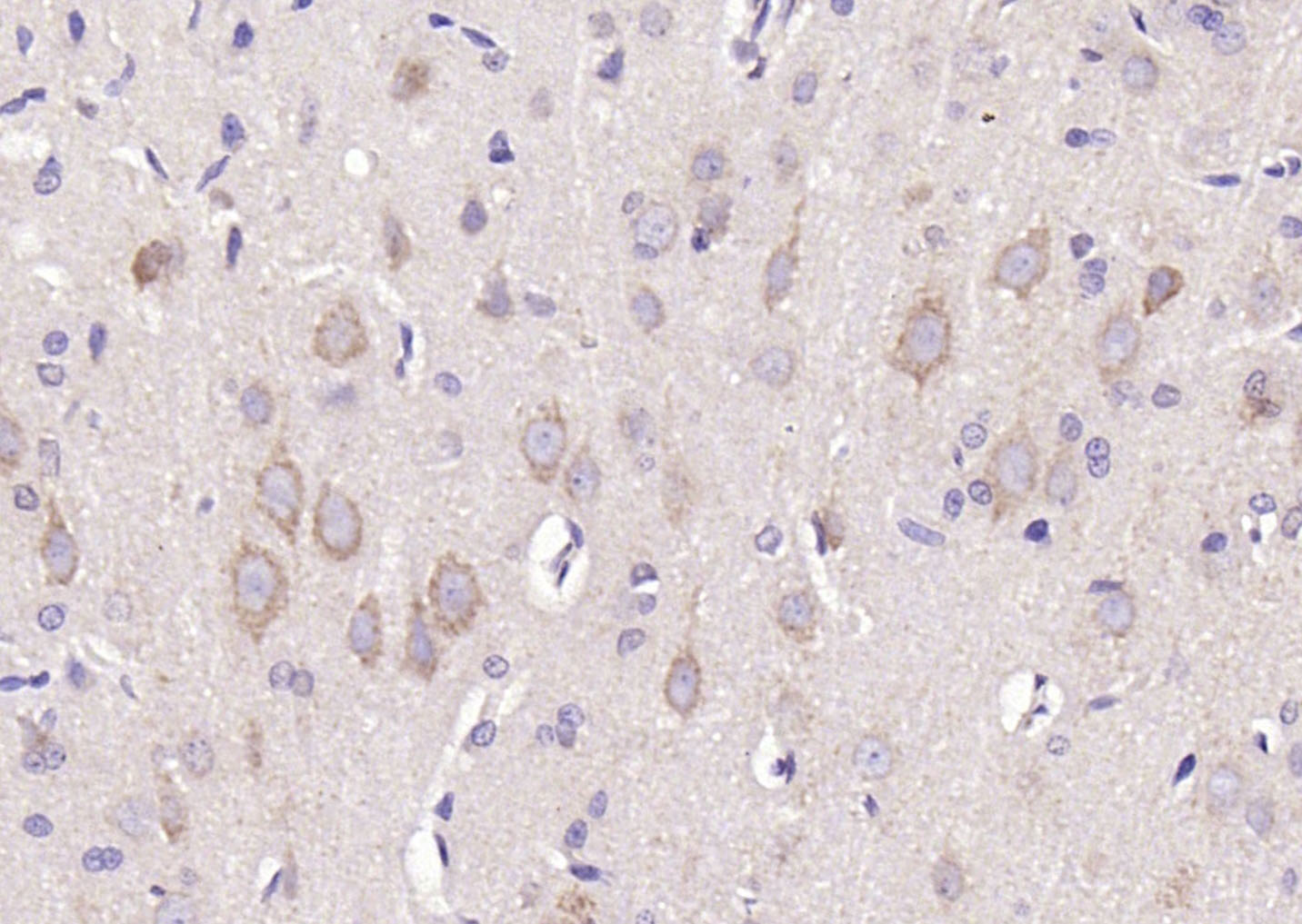 Paraformaldehyde-fixed, paraffin embedded (rat brain); Antigen retrieval by boiling in sodium citrate buffer (pH6.0) for 15min; Block endogenous peroxidase by 3% hydrogen peroxide for 20 minutes; Blocking buffer (normal goat serum) at 37°C for 30min; Antibody incubation with (PI 3 Kinase Class 3) Polyclonal Antibody, Unconjugated (SL4159R) at 1:200 overnight at 4°C, followed by operating according to SP Kit(Rabbit) (sp-0023) instructionsand DAB staining.
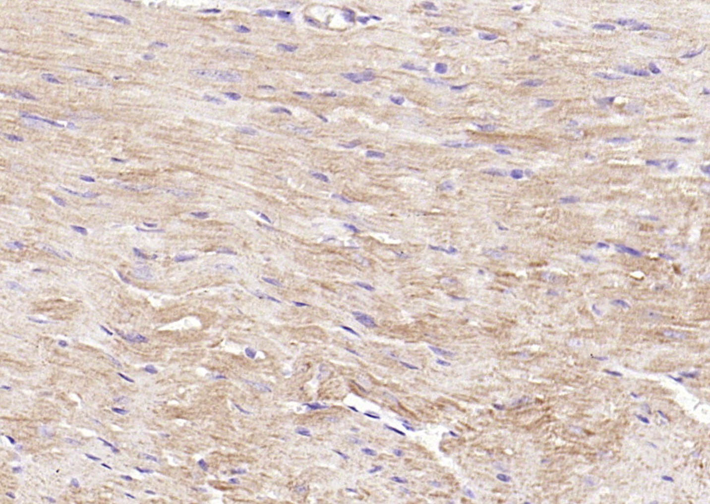 Paraformaldehyde-fixed, paraffin embedded (mouse heart ); Antigen retrieval by boiling in sodium citrate buffer (pH6.0) for 15min; Block endogenous peroxidase by 3% hydrogen peroxide for 20 minutes; Blocking buffer (normal goat serum) at 37°C for 30min; Antibody incubation with (PI 3 Kinase Class 3) Polyclonal Antibody, Unconjugated (SL4159R) at 1:200 overnight at 4°C, followed by operating according to SP Kit(Rabbit) (sp-0023) instructionsand DAB staining.
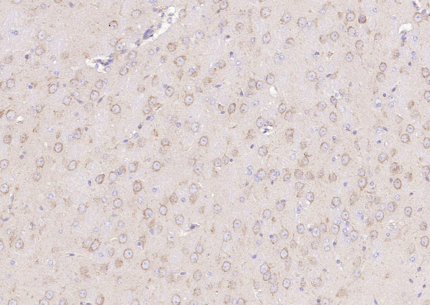 Paraformaldehyde-fixed, paraffin embedded (mouse brain); Antigen retrieval by boiling in sodium citrate buffer (pH6.0) for 15min; Block endogenous peroxidase by 3% hydrogen peroxide for 20 minutes; Blocking buffer (normal goat serum) at 37°C for 30min; Antibody incubation with (PI 3 Kinase Class 3) Polyclonal Antibody, Unconjugated (SL4159R) at 1:200 overnight at 4°C, followed by operating according to SP Kit(Rabbit) (sp-0023) instructionsand DAB staining.
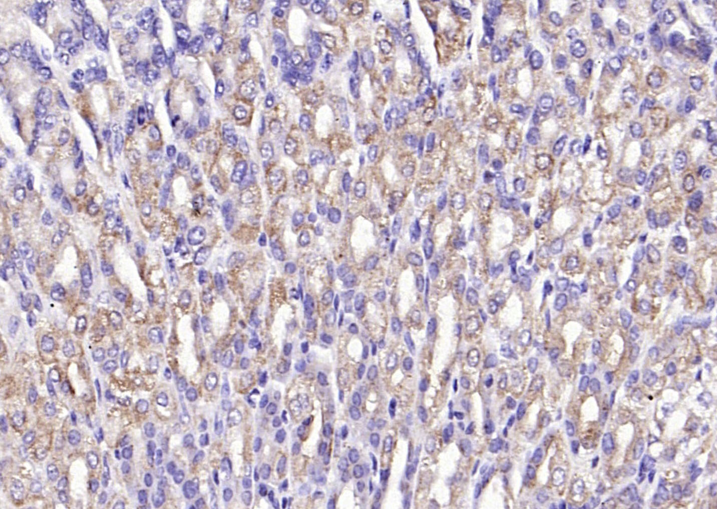 Paraformaldehyde-fixed, paraffin embedded (mouse stomach); Antigen retrieval by boiling in sodium citrate buffer (pH6.0) for 15min; Block endogenous peroxidase by 3% hydrogen peroxide for 20 minutes; Blocking buffer (normal goat serum) at 37°C for 30min; Antibody incubation with (PI 3 Kinase Class 3) Polyclonal Antibody, Unconjugated (SL4159R) at 1:200 overnight at 4°C, followed by operating according to SP Kit(Rabbit) (sp-0023) instructionsand DAB staining.
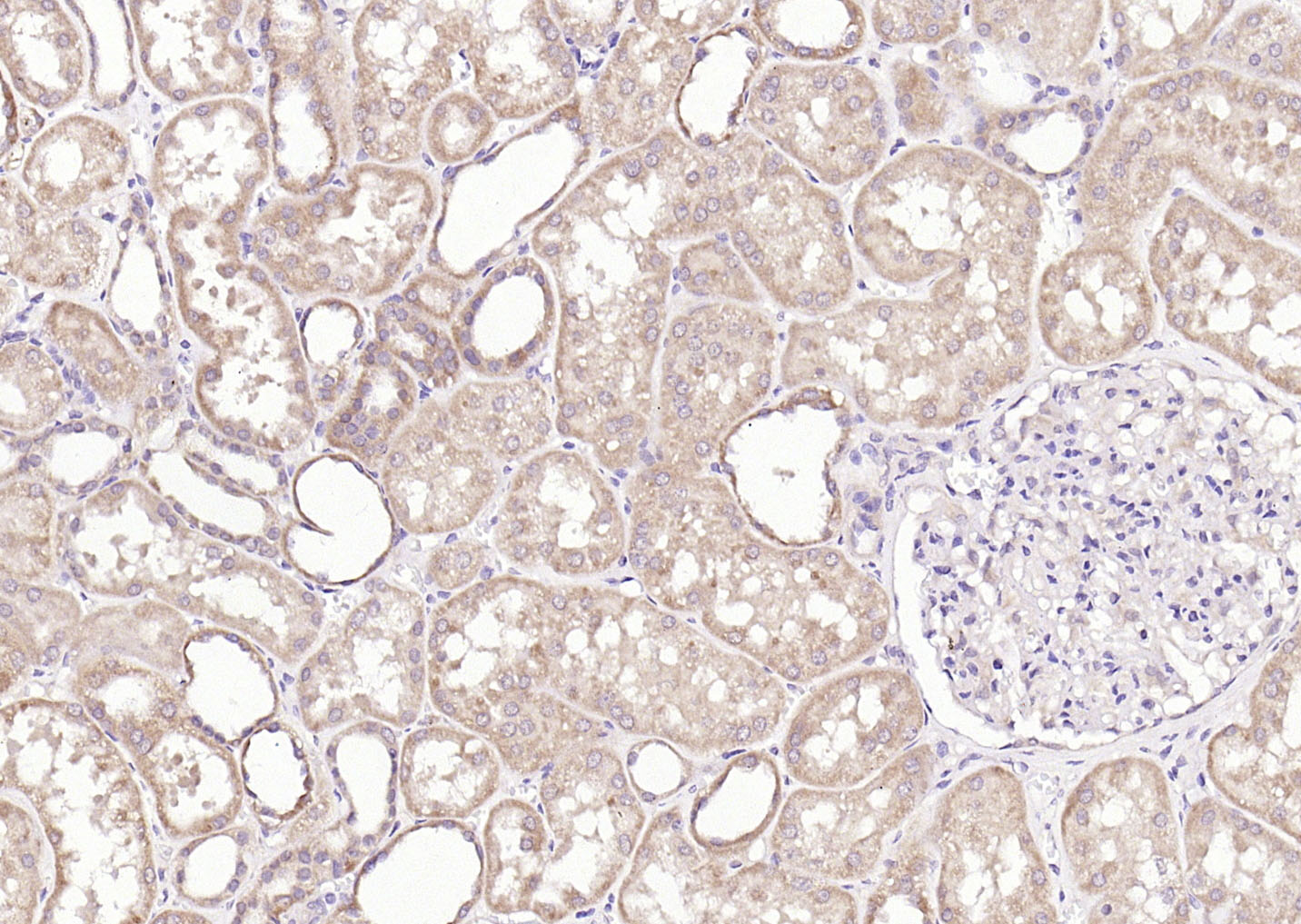 Paraformaldehyde-fixed, paraffin embedded (human kidney ); Antigen retrieval by boiling in sodium citrate buffer (pH6.0) for 15min; Block endogenous peroxidase by 3% hydrogen peroxide for 20 minutes; Blocking buffer (normal goat serum) at 37°C for 30min; Antibody incubation with (PI 3 Kinase Class 3) Polyclonal Antibody, Unconjugated (SL4159R) at 1:200 overnight at 4°C, followed by operating according to SP Kit(Rabbit) (sp-0023) instructionsand DAB staining.
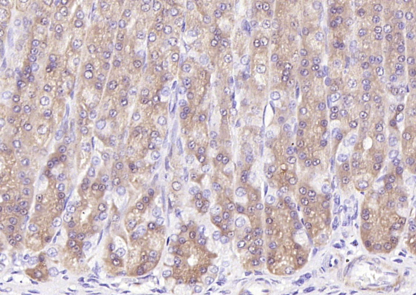 Paraformaldehyde-fixed, paraffin embedded (Rat stomach); Antigen retrieval by boiling in sodium citrate buffer (pH6.0) for 15min; Block endogenous peroxidase by 3% hydrogen peroxide for 20 minutes; Blocking buffer (normal goat serum) at 37°C for 30min; Antibody incubation with (PI 3 Kinase Class 3) Polyclonal Antibody, Unconjugated (SL4159R) at 1:200 overnight at 4°C, followed by operating according to SP Kit(Rabbit) (sp-0023) instructionsand DAB staining.
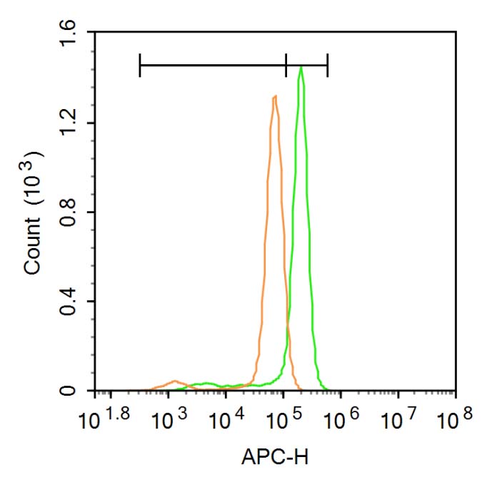 Blank control (Black line): HUVEC (Black).
Primary Antibody (green line): Rabbit Anti-PI 3 Kinase Class 3 antibody (SL4159R)
Dilution: 1μg /10^6 cells;
Isotype Control Antibody (orange line): Rabbit IgG .
Secondary Antibody (white blue line): Goat anti-rabbit IgG-AF647
Dilution: 1μg /test.
Protocol
The cells were fixed with 4% PFA (10min at room temperature)and then permeabilized with 0.1% PBST for 20 min at room temperature. The cells were then incubated in 5%BSA to block non-specific protein-protein interactions for 30 min at room temperature .Cells stained with Primary Antibody for 30 min at room temperature. The secondary antibody used for 40 min at room temperature. Acquisition of 20,000 events was performed.
Cartpieces
Totalgoods,subtotals:¥Checkout
References (0)
No References
Bought notes(bought amounts latest0)
No one bought this product
User Comment(Total0User Comment Num)
- No comment


 +86 571 56623320
 +86 18668110335

