
Rabbit Anti-IFNAR1 antibody
Interferon alpha/beta receptor 1; IFN-R-1; IFN-alpha/beta receptor 1; Cytokine receptor class-II member 1; Cytokine receptor family 2 member 1; CRF2-1; Type I interferon receptor 1; IFNAR; IFNAR 1; IFNAR-1; IFRC; Interferon alpha/beta receptor alpha chain
View History [Clear]
Details
Product Name IFNAR1 Chinese Name Interferonα/β受体1抗体 Alias Interferon alpha/beta receptor 1; IFN-R-1; IFN-alpha/beta receptor 1; Cytokine receptor class-II member 1; Cytokine receptor family 2 member 1; CRF2-1; Type I interferon receptor 1; IFNAR; IFNAR 1; IFNAR-1; IFRC; Interferon alpha/beta receptor alpha chain; INAR1_HUMAN. literatures Research Area Tumour immunology Chromatin and nuclear signals Signal transduction transcriptional regulatory factor Interferon Immunogen Species Rabbit Clonality Polyclonal React Species Human, Mouse, Rat, (predicted: Cow, Sheep, ) Applications WB=1:500-2000 ELISA=1:5000-10000 IHC-P=1:100-500 IHC-F=1:100-500 Flow-Cyt=1ug/test IF=1:100-500 (Paraffin sections need antigen repair)
not yet tested in other applications.
optimal dilutions/concentrations should be determined by the end user.Theoretical molecular weight 61kDa Cellular localization The cell membrane Form Liquid Concentration 1mg/ml immunogen KLH conjugated synthetic peptide derived from human IFNAR1: 351-450/557 <Extracellular> Lsotype IgG Purification affinity purified by Protein A Buffer Solution 0.01M TBS(pH7.4) with 1% BSA, 0.03% Proclin300 and 50% Glycerol. Storage Shipped at 4℃. Store at -20 °C for one year. Avoid repeated freeze/thaw cycles. Attention This product as supplied is intended for research use only, not for use in human, therapeutic or diagnostic applications. PubMed PubMed Product Detail IFNAR1 is a member of the cytokine receptor superfamily which also includes receptors for interleukins, IFN gamma, ciliary neurotrophic factor, somatotrophin, erythropoietin, nerve growth factor, tumor necrosis factor, leukemia inhibitory factor, and oncostatin M. Some members of the family have an alpha chain with either low or high ligand binding affinity and at least one beta chain involved in signal transduction with either relatively low or no ligand binding affinity. Type I interferons, alpha and beta, induce a variety of effects on target cells including antiviral, antiproliferative, and immunomodulatory activities. The alpha and beta interferons compete to bind to a common cell surface receptor, while IFN gamma binds to a distinct receptor. IFNAR1 is very responsive to type I interferons and bind to IFN beta and IFN alpha subtypes. It is also functionally involved in signal transduction because of its association with the cytoplasmic tyrosine kinase JAK1. The type I interferons, alpha and beta, are produced by leukocytes (alpha subunits), fibroblasts (beta subtypes), lymphocytes (omega subtypes), and ruminant embryos (tau subtypes). Interferon receptors are generally found on most human cell types whatever their origin, even on cells poorly responsive to interferon. IFNAR1 is expressed on the cell surface in a variety of human cell lines.
Function:
Associates with IFNAR2 to form the type I interferon receptor. Receptor for interferons alpha and beta. Binding to type I IFNs triggers tyrosine phosphorylation of a number of proteins including JAKs, TYK2, STAT proteins and IFNR alpha- and beta-subunits themselves.
Subunit:
Heterodimer with IFNAR2; in presence of interferon alpha and beta ligands, the heterodimer forms the type I interferon receptor. Interacts with STAT1 and STAT2. Interacts with IFNAR2.
Subcellular Location:
Membrane; Single-pass type I membrane protein.
Post-translational modifications:
Phosphorylated on tyrosine residues by TYK2 tyrosine kinase.
Palmitoylation at Cys-463 is required for the activation of STAT1 and STAT2.
Similarity:
Belongs to the type II cytokine receptor family.
Contains 3 fibronectin type-III domains.
SWISS:
P17181
Gene ID:
3454
Database links:Entrez Gene: 3454 Human
Entrez Gene: 15975 Mouse
Omim: 107450 Human
SwissProt: P17181 Human
SwissProt: P33896 Mouse
Unigene: 529400 Human
Unigene: 502 Mouse
Product Picture 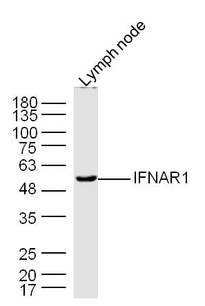 Sample: Lymph node (Mouse) Lysate at 40 ug
Primary: Anti- IFNAR1 (SL4116R) at 1/300 dilution
Secondary: IRDye800CW Goat Anti-Rabbit IgG at 1/20000 dilution
Predicted band size: 61 kD
Observed band size: 61 kD
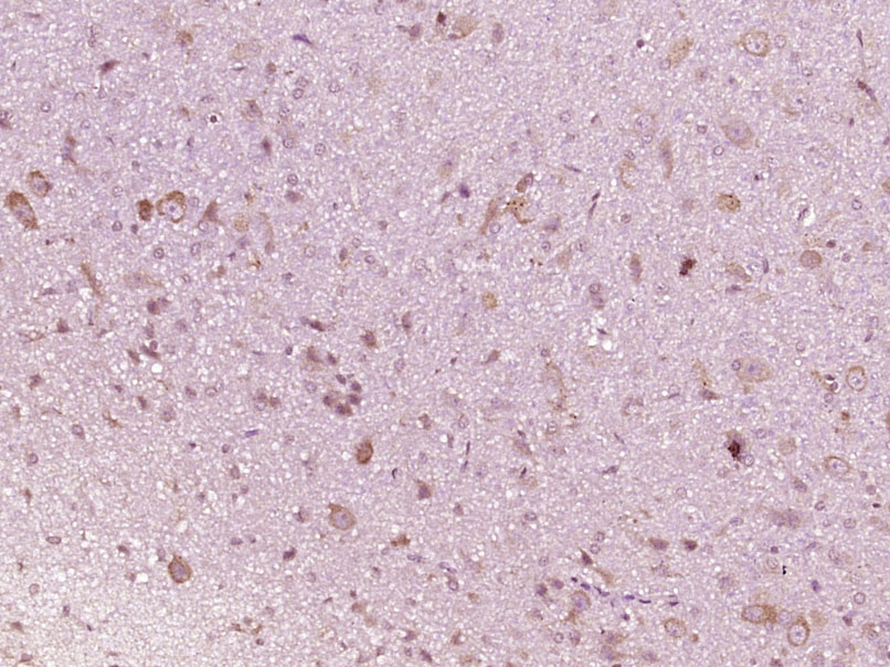 Paraformaldehyde-fixed, paraffin embedded (Rat brain); Antigen retrieval by boiling in sodium citrate buffer (pH6.0) for 15min; Block endogenous peroxidase by 3% hydrogen peroxide for 20 minutes; Blocking buffer (normal goat serum) at 37°C for 30min; Antibody incubation with (IFNAR1) Polyclonal Antibody, Unconjugated (SL4116R) at 1:400 overnight at 4°C, followed by operating according to SP Kit(Rabbit) (sp-0023) instructionsand DAB staining.
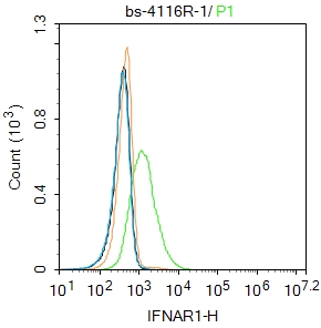 Blank control:K562.
Primary Antibody (green line): Rabbit Anti-IFNAR1 antibody (SL4116R)
Dilution: 1ug/Test;
Secondary Antibody : Goat anti-rabbit IgG-FITC
Dilution: 0.5ug/Test.
Protocol
The cells were incubated in 5%BSA to block non-specific protein-protein interactions for 30 min at room temperature .Cells stained with Primary Antibody for 30 min at room temperature. The secondary antibody used for 40 min at room temperature. Acquisition of 20,000 events was performed.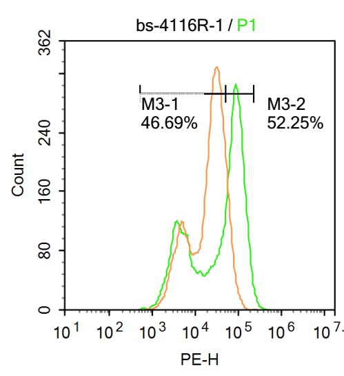 Blank control:U-2OS.
Primary Antibody (green line): Rabbit Anti-TNNT2 antibody (SL4116R)
Dilution: 1μg /10^6 cells;
Isotype Control Antibody (orange line): Rabbit IgG .
Secondary Antibody : Goat anti-rabbit IgG-AF647
Dilution: 1μg /test.
Protocol
The cells were fixed with 4% PFA (10min at room temperature)and then permeabilized with 0.1% PBST for 20 min at room temperature. The cells were then incubated in 5%BSA to block non-specific protein-protein interactions for 30 min at at room temperature .Cells stained with Primary Antibody for 30 min at room temperature. The secondary antibody used for 40 min at room temperature. Acquisition of 20,000 events was performed.
Cartpieces
Totalgoods,subtotals:¥Checkout
Bought notes(bought amounts latest0)
No one bought this product
User Comment(Total0User Comment Num)
- No comment


 +86 571 56623320
 +86 18668110335

