
Rabbit Anti-G6PC antibody
glucose-6-phosphatase, catalytic subunit; GSD1; AW107337; G-6-Pase; G6Pase; G6Pase-alpha; Glucose 6 phosphatase alpha; G6PC_HUMAN; G6PT; Glucose-6-phosphatase alpha; Glucose-6-phosphatase; GSD1a; MGC163350; MGC93613; RP23-281C18.19.
View History [Clear]
Details
Product Name G6PC Chinese Name 葡萄糖6磷酸酶α/G6Pase-α抗体 Alias glucose-6-phosphatase, catalytic subunit; GSD1; AW107337; G-6-Pase; G6Pase; G6Pase-alpha; Glucose 6 phosphatase alpha; G6PC_HUMAN; G6PT; Glucose-6-phosphatase alpha; Glucose-6-phosphatase; GSD1a; MGC163350; MGC93613; RP23-281C18.19. literatures Research Area Tumour immunology transcriptional regulatory factor Kinases and Phosphatases Immunogen Species Rabbit Clonality Polyclonal React Species Human, Rat, (predicted: Mouse, Dog, Pig, Cow, Rabbit, Sheep, ) Applications WB=1:500-2000 ELISA=1:5000-10000 IHC-P=1:100-500 Flow-Cyt=0.2ug/test (Paraffin sections need antigen repair)
not yet tested in other applications.
optimal dilutions/concentrations should be determined by the end user.Theoretical molecular weight 39kDa Cellular localization cytoplasmic The cell membrane Form Liquid Concentration 1mg/ml immunogen KLH conjugated synthetic peptide derived from human Glucose 6 phosphatase alpha: 81-180/357 Lsotype IgG Purification affinity purified by Protein A Buffer Solution 0.01M TBS(pH7.4) with 1% BSA, 0.03% Proclin300 and 50% Glycerol. Storage Shipped at 4℃. Store at -20 °C for one year. Avoid repeated freeze/thaw cycles. Attention This product as supplied is intended for research use only, not for use in human, therapeutic or diagnostic applications. PubMed PubMed Product Detail Glucose-6-phosphatase (G6Pase) is a multi-subunit integral membrane protein of the endoplasmic reticulum that is composed of a catalytic subunit and transporters for G6P, inorganic phosphate, and glucose. This gene (G6PC) is one of the three glucose-6-phosphatase catalytic-subunit-encoding genes in human: G6PC, G6PC2 and G6PC3. Glucose-6-phosphatase catalyzes the hydrolysis of D-glucose 6-phosphate to D-glucose and orthophosphate and is a key enzyme in glucose homeostasis, functioning in gluconeogenesis and glycogenolysis. Mutations in this gene cause glycogen storage disease type I (GSD1). This disease, also known as von Gierke disease, is a metabolic disorder characterized by severe hypoglycemia associated with the accumulation of glycogen and fat in the liver and kidneys.[provided by RefSeq, Feb 2011]
Function:
Hydrolyzes glucose-6-phosphate to glucose in the endoplasmic reticulum. Forms with the glucose-6-phosphate transporter (SLC37A4/G6PT) the complex responsible for glucose production through glycogenolysis and gluconeogenesis. Hence, it is the key enzyme in homeostatic regulation of blood glucose levels.
Subcellular Location:
Endoplasmic reticulum membrane; Multi-pass membrane protein.
DISEASE:
Defects in G6PC are the cause of glycogen storage disease type 1A (GSD1A) [MIM:232200]. A metabolic disorder characterized by impairment of terminal steps of glycogenolysis and gluconeogenesis. Patients manifest a wide range of clinical symptoms and biochemical abnormalities, including hypoglycemia, severe hepatomegaly due to excessive accumulation of glycogen, kidney enlargement, growth retardation, lactic acidemia, hyperlipidemia, and hyperuricemia.
Similarity:
Belongs to the glucose-6-phosphatase family.
SWISS:
P35575
Gene ID:
2538
Database links:
Entrez Gene: 2538 Human
Entrez Gene: 14377 Mouse
SwissProt: P35575 Human
SwissProt: P35576 Mouse
Product Picture 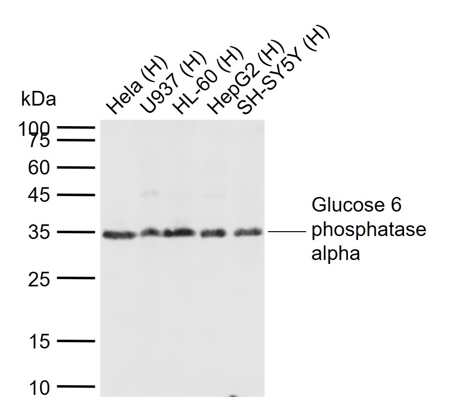 Sample:
Lane 1: Human Hela cell lysates
Lane 2: Human U937cell lysates
Lane 3: Human HL-60 cell lysates
Lane 4: Human HepG2 cell lysates
Lane 5: Human SH-SY5Y cell lysates
Primary: Anti-Glucose 6 phosphatase alpha (SL4044R) at 1/1000 dilution
Secondary: IRDye800CW Goat Anti-Rabbit IgG at 1/20000 dilution
Predicted band size: 39 kDa
Observed band size: 35 kDa
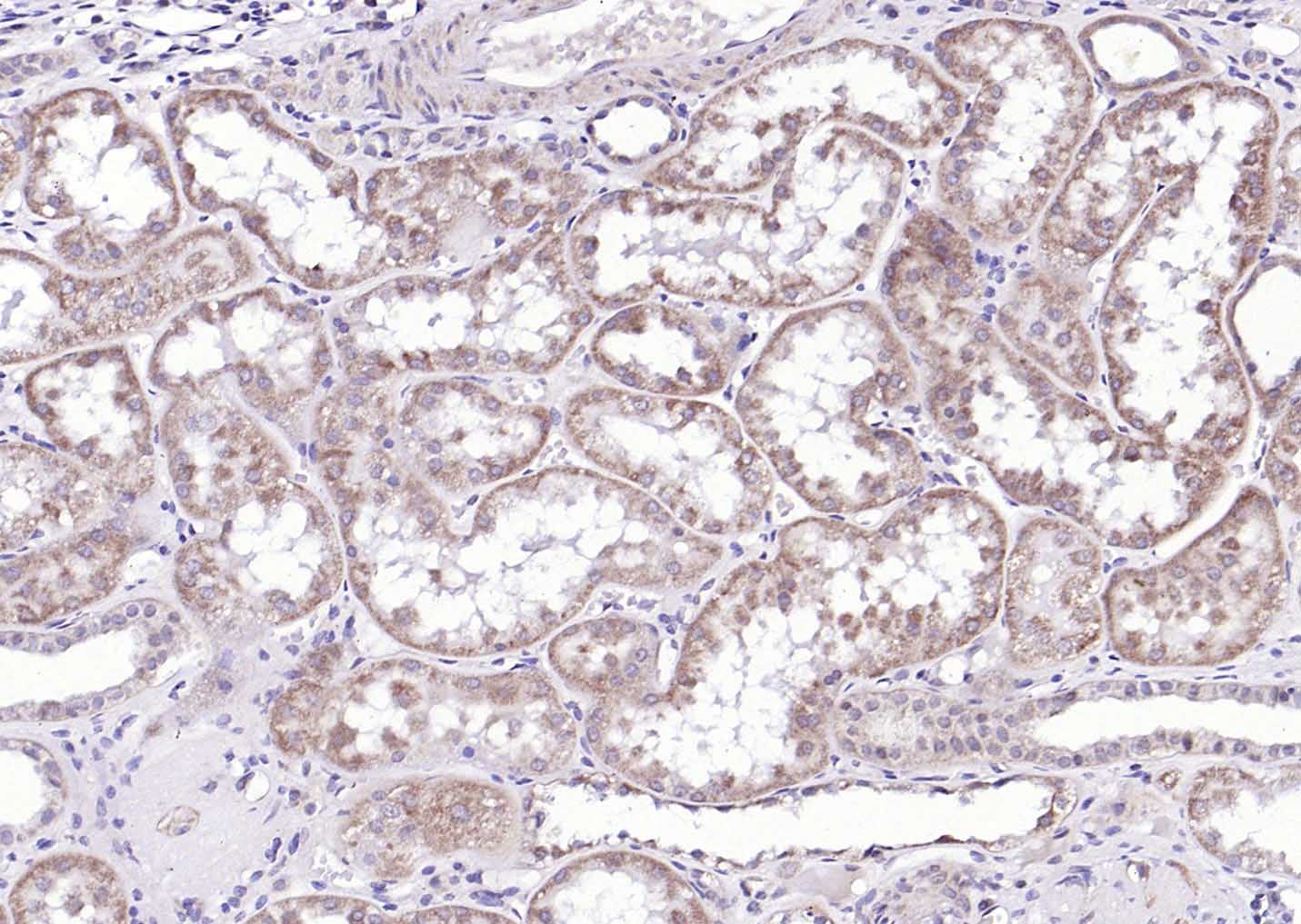 Paraformaldehyde-fixed, paraffin embedded (Human kidney); Antigen retrieval by boiling in sodium citrate buffer (pH6.0) for 15min; Block endogenous peroxidase by 3% hydrogen peroxide for 20 minutes; Blocking buffer (normal goat serum) at 37°C for 30min; Antibody incubation with (Glucose 6 phosphatase alpha) Polyclonal Antibody, Unconjugated (SL4044R) at 1:200 overnight at 4°C, followed by operating according to SP Kit(Rabbit) (sp-0023) instructionsand DAB staining.
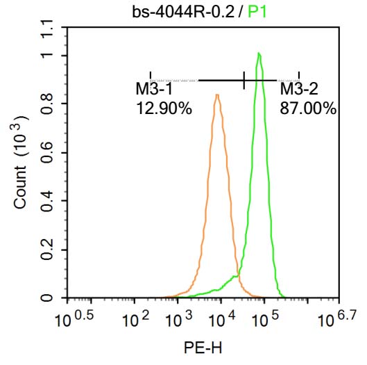 U-937 cells were incubated in 5% BSA blocking buffer for 30 min at room temperature. Cells were then stained with SL4044R Antibody at 1:500 dilution in blocking buffer and incubated for 30 min at room temperature, washed twice with 2%BSA in PBS, followed by secondary antibody incubation for 40 min at room temperature. Acquisitions of 20,000 events were performed. Cells stained with primary antibody (green), and isotype control (orange).U-937 cells were incubated in 5% BSA blocking buffer for 30 min at room temperature. Cells were then stained with SL4044R Antibody at 1:500 dilution in blocking buffer and incubated for 30 min at room temperature, washed twice with 2%BSA in PBS, followed by secondary antibody incubation for 40 min at room temperature. Acquisitions of 20,000 events were performed. Cells stained with primary antibody (green), and isotype control (orange).
Cartpieces
Totalgoods,subtotals:¥Checkout
References (0)
No References
Bought notes(bought amounts latest0)
No one bought this product
User Comment(Total0User Comment Num)
- No comment


 +86 571 56623320
 +86 18668110335

