
Rabbit Anti-Synaptopodin antibody
9030217H17RIK; 9130229N11; 9330140I15Rik; AW046661; SYNAPTOPODIN; KIAA1029; SYNPO; SYNPO_HUMAN.
View History [Clear]
Details
Product Name Synaptopodin Chinese Name 突触足蛋白抗体 Alias 9030217H17RIK; 9130229N11; 9330140I15Rik; AW046661; SYNAPTOPODIN; KIAA1029; SYNPO; SYNPO_HUMAN. literatures Research Area Tumour Cell biology immunology Neurobiology Signal transduction Apoptosis transcriptional regulatory factor Immunogen Species Rabbit Clonality Polyclonal React Species Human, Mouse, Rat, Applications WB=1:500-2000 IHC-P=1:100-500 Flow-Cyt=1μg /test (Paraffin sections need antigen repair)
not yet tested in other applications.
optimal dilutions/concentrations should be determined by the end user.Theoretical molecular weight 102kDa Cellular localization cytoplasmic The cell membrane Form Liquid Concentration 1mg/ml immunogen KLH conjugated synthetic peptide derived from human Synaptopodin: 601-700/903 Lsotype IgG Purification affinity purified by Protein A Buffer Solution 0.01M TBS(pH7.4) with 1% BSA, 0.03% Proclin300 and 50% Glycerol. Storage Shipped at 4℃. Store at -20 °C for one year. Avoid repeated freeze/thaw cycles. Attention This product as supplied is intended for research use only, not for use in human, therapeutic or diagnostic applications. PubMed PubMed Product Detail Synaptopodin is an actin-associated protein that may play a role in actin-based cell shape and motility. May be essential for the formation of spine apparatuses in spines of telencephalic neurons, involved in synaptic plasticity. The name synaptopodin derives from the protein's associations with postsynaptic densities and dendritic spines and with renal podocytes.
Subunit:
Interacts with BAIAP1. Interacts with actin (By similarity). Interacts (via PPxY motifs) with WWC1 (via WW domains).
Subcellular Location:
Cytoplasm, cytoskeleton (By similarity). Cell junction, tight junction (By similarity). Perikaryon (By similarity). Cell projection, dendritic spine (By similarity). Cell junction, synapse, postsynaptic cell membrane, postsynaptic density (By similarity). Cell junction, synapse (By similarity). Note=Localized at the tight junction of cells. In brain, localized to the postsynaptic densities and in the perikarya. Asssociated with dendritic spines of a subset of synapses (By similarity).
Tissue Specificity:
Expressed in brain, namely in the olfactory bulb, cerebral cortex, striatum, and hippocampus, but not in the cerebellum. Also expressed in the podocytes of kidney glomeruli. In the hippocampus, mainly expressed in the principal cell layer of the dentate gyrus and Ammon's horn.
Similarity:
Belongs to the synaptopodin family.
SWISS:
Q8N3V7
Gene ID:
11346
Database links:Entrez Gene: 11346 Human
Entrez Gene: 104027 Mouse
Omim: 608155 Human
SwissProt: Q8N3V7 Human
SwissProt: Q8CC35 Mouse
Unigene: 435228 Human
synaptopodin是一种与肌动蛋白微丝偶联的蛋白,其富含脯氨酸,为线状蛋白质,在动物体内表达于肾小球的足细胞和后脑的突触内,synaptopodin被认为是足细胞特异的分化成熟Maker。Product Picture 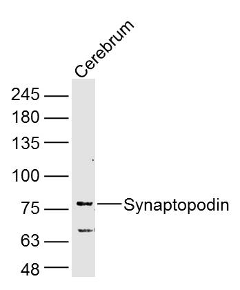 Sample: Cerebrum (Rat) Lysate at 40 ug
Primary: Anti-Synaptopodin (SL3633R) at 1/300 dilution
Secondary: IRDye800CW Goat Anti-Rabbit IgG at 1/20000 dilution
Predicted band size: 102 kD
Observed band size: 76 kD
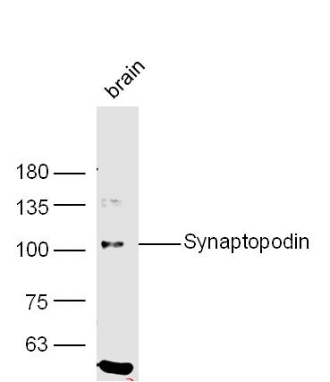 Sample: Brain (Mouse) Lysate at 40 ug
Primary: Anti-Synaptopodin (SL3633R) at 1/300 dilution
Secondary: IRDye800CW Goat Anti-Rabbit IgG at 1/20000 dilution
Predicted band size: 102 kD
Observed band size: 102 kD
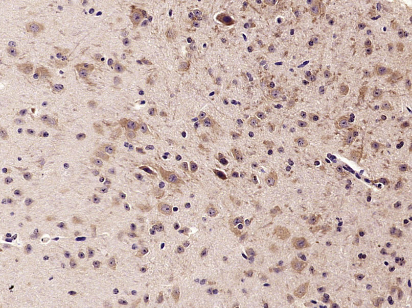 Paraformaldehyde-fixed, paraffin embedded (Mouse brain); Antigen retrieval by microwave in sodium citrate buffer (pH6.0) ; Block endogenous peroxidase by 3% hydrogen peroxide for 30 minutes; Blocking buffer (3% BSA) at RT for 30min; Antibody incubation with (Synaptopodin) Polyclonal Antibody, Unconjugated (SL3633R) at 1:400 overnight at 4°C, followed by conjugation to the secondary antibody (labeled with HRP)and DAB staining.
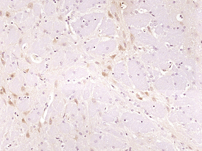 Paraformaldehyde-fixed, paraffin embedded (Rat brain); Antigen retrieval by boiling in sodium citrate buffer (pH6.0) for 15min; Block endogenous peroxidase by 3% hydrogen peroxide for 20 minutes; Blocking buffer (normal goat serum) at 37°C for 30min; Antibody incubation with (Synaptopodin) Polyclonal Antibody, Unconjugated (SL3633R) at 1:400 overnight at 4°C, followed by operating according to SP Kit(Rabbit) (sp-0023) instructionsand DAB staining.
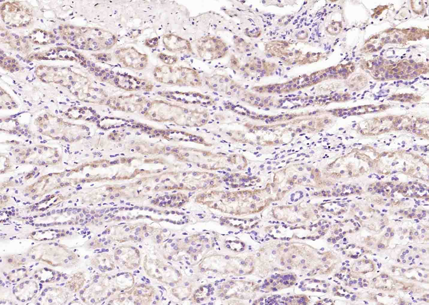 Paraformaldehyde-fixed, paraffin embedded (Human kidney); Antigen retrieval by boiling in sodium citrate buffer (pH6.0) for 15min; Block endogenous peroxidase by 3% hydrogen peroxide for 20 minutes; Blocking buffer (normal goat serum) at 37°C for 30min; Antibody incubation with (Synaptopodin) Polyclonal Antibody, Unconjugated (SL3633R) at 1:200 overnight at 4°C, followed by operating according to SP Kit(Rabbit) (sp-0023) instructionsand DAB staining.
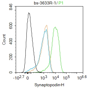 Blank control(black line):SH-SY5Y.
Primary Antibody (green line): Rabbit Anti-Synaptopodin antibody (SL3633R)
Dilution:1ug/Test;
Secondary Antibody(white blue line): Goat anti-rabbit IgG-AF488
Dilution: 0.5ug/Test.
Isotype control(orange line): Normal Rabbit IgG
Protocol
The cells were fixed with 4% PFA (10min at room temperature)and then permeabilized with 90% ice-cold methanol for 20 min at -20℃, The cells were then incubated in 5%BSA to block non-specific protein-protein interactions for 30 min at room temperature .Cells stained with Primary Antibody for 30 min at room temperature. The secondary antibody used for 40 min at room temperature. Acquisition of 20,000 events was performed. Positive control: RSC96
Isotype Control Antibody: Rabbit IgG; Secondary Antibody: Goat anti-rabbit IgG-FITC; Dilution: 1:200 in 1 X PBS containing 0.5% BSA
Primary Antibody catalog number: SL3633R; Dilution: 1μg in 100 μl 1X PBS containing 0.5% BSA
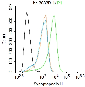 Blank control(black line):SH-SY5Y.
Primary Antibody (green line): Rabbit Anti-Synaptopodin antibody (SL3633R)
Dilution:1ug/Test;
Secondary Antibody(white blue line): Goat anti-rabbit IgG-AF488
Dilution: 0.5ug/Test.
Isotype control(orange line): Normal Rabbit IgG
Protocol
The cells were fixed with 4% PFA (10min at room temperature)and then permeabilized with 90% ice-cold methanol for 20 min at -20℃, The cells were then incubated in 5%BSA to block non-specific protein-protein interactions for 30 min at room temperature .Cells stained with Primary Antibody for 30 min at room temperature. The secondary antibody used for 40 min at room temperature. Acquisition of 20,000 events was performed.
Cartpieces
Totalgoods,subtotals:¥Checkout
References (0)
No References
Bought notes(bought amounts latest0)
No one bought this product
User Comment(Total0User Comment Num)
- No comment


 +86 571 56623320
 +86 18668110335

