
Rabbit Anti-phospho-Cyclin E1 (Thr77)antibody
Cyclin E1 (phospho-Thr77); Cyclin E1 (phospho-T77); CCNE 1; CCNE; CCNE1; Cyclin Es; Cyclin Et; CyclinE; G1/S specific cyclin E; G1/S-specific cyclin-E1; CCNE1_HUMAN; pCCNE1.
View History [Clear]
Details
Product Name phospho-Cyclin E1 (Thr77) Chinese Name 磷酸化周期素E1抗体 Alias Cyclin E1 (phospho-Thr77); Cyclin E1 (phospho-T77); CCNE 1; CCNE; CCNE1; Cyclin Es; Cyclin Et; CyclinE; G1/S specific cyclin E; G1/S-specific cyclin-E1; CCNE1_HUMAN; pCCNE1. Product Type Phosphorylated anti Research Area Tumour Cell biology Cyclin Immunogen Species Rabbit Clonality Polyclonal React Species Human, (predicted: Mouse, Rat, Chicken, Dog, Pig, Cow, Horse, Rabbit, ) Applications WB=1:500-2000 ELISA=1:5000-10000 Flow-Cyt=1μg /Test
not yet tested in other applications.
optimal dilutions/concentrations should be determined by the end user.Theoretical molecular weight 47kDa Cellular localization The nucleus Form Liquid Concentration 1mg/ml immunogen KLH conjugated Synthesised phosphopeptide derived from human Cyclin E around the phosphorylation site of Thr77: IP(p-T)PD Lsotype IgG Purification affinity purified by Protein A Buffer Solution 0.01M TBS(pH7.4) with 1% BSA, 0.03% Proclin300 and 50% Glycerol. Storage Shipped at 4℃. Store at -20 °C for one year. Avoid repeated freeze/thaw cycles. Attention This product as supplied is intended for research use only, not for use in human, therapeutic or diagnostic applications. PubMed PubMed Product Detail Cyclin E is a regulatory subunit of Cdk2 and controls G1 / S transition during the mammalian cell cycle. Multiple isoforms of Cyclin E are only expressed in tumors but not in normal tissue, suggesting a post transcriptional regulation of Cyclin E. In vitro analyses indicated that these truncated variant isoforms of Cyclin E are able to phosphorylate histone H1. Alterations in the Cyclin E protein have been implicated as indicators of worse prognosis in various cancers.
Function:
Essential for the control of the cell cycle at the G1/S (start) transition.
Subunit:
Interacts with a member of the CDK2/CDK protein kinases to form a serine/threonine kinase holoenzyme complex. The cyclin subunit imparts substrate specificity to the complex. Found in a complex with CDK2, CABLES1 and CCNA1 (By similarity). Part of a complex consisting of UHRF2, CDK2 and CCNE1. Interacts directly with UHRF2; the interaction ubiquitinates CCNE1 and appears to occur independently of CCNE1 phosphorylation.
Subcellular Location:
Nucleus.
Tissue Specificity:
Highly expressed in testis and placenta. Low levels in bronchial epithelial cells.
Post-translational modifications:
Phosphorylation of Thr-395 by GSK3 and of Ser-399 by CDK2 accelerates degradation via the ubiquitin proteasome pathway. Phosphorylated upon DNA damage, probably by ATM or ATR.
Ubiquitinated by UHRF2; appears to occur independently of phosphorylation.
Similarity:
Belongs to the cyclin family. Cyclin E subfamily.
SWISS:
P24864
Gene ID:
898
Database links:Entrez Gene: 898 Human
Entrez Gene: 12447 Mouse
Omim: 123837 Human
SwissProt: P24864 Human
SwissProt: Q61457 Mouse
Unigene: 244723 Human
Unigene: 16110 Mouse
Unigene: 15455 Rat
细胞周期素E是调控细胞G-1→S期转变的关键因素。由于在多种Tumour中的不适当表达,细胞周期素E现在已明确为原癌基因。Product Picture 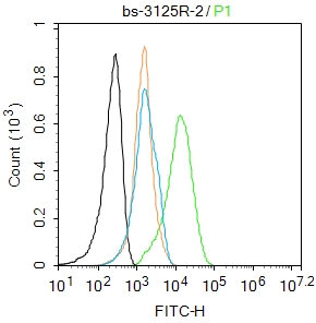 Blank control:Hela.
Primary Antibody (green line): Rabbit Anti-phospho-Cyclin E1 (Thr77) antibody (SL3152R)
Dilution: 2μg /10^6 cells;
Isotype Control Antibody (orange line): Rabbit IgG .
Secondary Antibody : Goat anti-rabbit IgG-FITC
Dilution: 1μg /test.
Protocol
The cells were fixed with 4% PFA (10min at room temperature)and then permeabilized with 0.1%PBST for 20 min at room temperature. The cells were then incubated in 5%BSA to block non-specific protein-protein interactions for 30 min at room temperature .Cells stained with Primary Antibody for 30 min at room temperature. The secondary antibody used for 40 min at room temperature. Acquisition of 20,000 events was performed.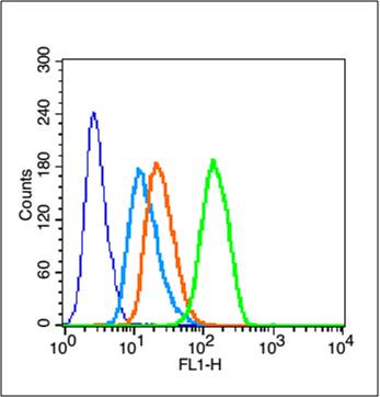 Blank control (blue line): MCF7(fixed with 70% ethanol (Overnight at 4℃) and then permeabilized with 90% ice-cold methanol for 30 min on ice)
Primary Antibody (green line): Rabbit Anti-phospho-Cyclin E1 (Thr77) antibody (SL3125R),Dilution: 3μg /10^6 cells.
Isotype Control Antibody (orange line): Rabbit IgG .
Secondary Antibody (white blue line): Goat anti-rabbit IgG-FITC,Dilution: 1μg /test.
The cells were . Cells stained with Primary Antibody for 30 min at room temperature. The cells were then incubated in 1 X PBS/2%BSA/10% goat serum to block non-specific protein-protein interactions followed by the antibody for 15 min at room temperature. The secondary antibody used for 40 min at room temperature. Acquisition of 20,000 events was performed.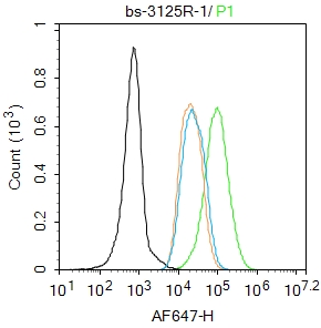 Blank control: MCF7.
Primary Antibody (green line): Rabbit Anti-phospho-Cyclin E1 (Thr77) antibody (SL3125R)
Dilution: 2μg /10^6 cells;
Isotype Control Antibody (orange line): Rabbit IgG .
Secondary Antibody : Goat anti-rabbit IgG-AF647
Dilution: 1μg /test.
Protocol
The cells were fixed with 4% PFA (10min at room temperature)and then permeabilized with 90% ice-cold methanol for 20 min at-20℃.The cells were then incubated in 5%BSA to block non-specific protein-protein interactions for 30 min at room temperature .Cells stained with Primary Antibody for 30 min at room temperature. The secondary antibody used for 40 min at room temperature. Acquisition of 20,000 events was performed.Blank control: MCF7.
Primary Antibody (green line): Rabbit Anti-phospho-Cyclin E1 (Thr77) antibody (SL3125R)
Dilution: 2μg /10^6 cells;
Isotype Control Antibody (orange line): Rabbit IgG .
Secondary Antibody : Goat anti-rabbit IgG-AF647
Dilution: 1μg /test.
Protocol
The cells were fixed with 4% PFA (10min at room temperature)and then permeabilized with 90% ice-cold methanol for 20 min at-20℃.The cells were then incubated in 5%BSA to block non-specific protein-protein interactions for 30 min at room temperature .Cells stained with Primary Antibody for 30 min at room temperature. The secondary antibody used for 40 min at room temperature. Acquisition of 20,000 events was performed.
Cartpieces
Totalgoods,subtotals:¥Checkout
Bought notes(bought amounts latest0)
No one bought this product
User Comment(Total0User Comment Num)
- No comment


 +86 571 56623320
 +86 18668110335

