
Rabbit Anti-JNK1+JNK2+JNK3 antibody
JNK1 + JNK2 + JNK3; JNK1/2/3; JNK1+2+3; JNK1 + JNK2 + JNK3; MAPK10; c Jun N terminal kinase 1; c Jun N terminal kinase 2; c Jun N terminal kinase 3; JNK; JNK1; JNK2; JNK2ALPHA; JNK2BETA; JNK3; MAPK8; MAPK9; Mitogen activated protein kinase 10; Mitogen act
View History [Clear]
Details
Product Name JNK1+JNK2+JNK3 Chinese Name 氨基末端激酶1/2/3抗体 Alias JNK1 + JNK2 + JNK3; JNK1/2/3; JNK1+2+3; JNK1 + JNK2 + JNK3; MAPK10; c Jun N terminal kinase 1; c Jun N terminal kinase 2; c Jun N terminal kinase 3; JNK; JNK1; JNK2; JNK2ALPHA; JNK2BETA; JNK3; MAPK8; MAPK9; Mitogen activated protein kinase 10; Mitogen activated protein kinase 8; Mitogen activated protein kinase 9; SAPK(beta); Stress activated protein kinase JNK1; Stress activated protein kinase JNK2; Stress activated protein kinase JNK3; SAPK; p54a; JNK2A; JNK2B; PRKM9; JNK-55; SAPK1a; JNK2BETA; p54aSAPK; JNK2ALPHA. literatures Research Area Cell biology Apoptosis Diabetes Immunogen Species Rabbit Clonality Polyclonal React Species Human, Mouse, Rat, (predicted: Dog, Pig, Cow, ) Applications WB=1:500-2000 ELISA=1:5000-10000 IHC-P=1:100-500 IHC-F=1:100-500 Flow-Cyt=1ug/Test ICC=1:100 IF=1:100-500 (Paraffin sections need antigen repair)
not yet tested in other applications.
optimal dilutions/concentrations should be determined by the end user.Theoretical molecular weight 42-47kDa Cellular localization The nucleus cytoplasmic Form Liquid Concentration 1mg/ml immunogen KLH conjugated synthetic peptide derived from human JNK1/2/3: 151-250/384 Lsotype IgG Purification affinity purified by Protein A Buffer Solution 0.01M TBS(pH7.4) with 1% BSA, 0.03% Proclin300 and 50% Glycerol. Storage Shipped at 4℃. Store at -20 °C for one year. Avoid repeated freeze/thaw cycles. Attention This product as supplied is intended for research use only, not for use in human, therapeutic or diagnostic applications. PubMed PubMed Product Detail JNK1(MAPK8) is a member of the MAP kinase family. JNK1 is activated by threonine and tyrosine phosphorylation by either of two dual specificity kinases, MAP2K4 and MAP2K7.
JNK2 (p54a, SAPK1a), along with JNK1 and JNK3, is thought to play an important role in nuclear signal transduction through its environmental stress activation and subsequent phosphorylation of the nuclear transcription factor p53.
JNK3 is a neuron-specific form of c-Jun N-terminal kinases. Through its phosphorylation and nuclear localization, this kinase plays regulatory roles in the signaling pathways of neuronal apoptosis.
The JNK pathway is critically involved in diabetes and levels are abnormally elevated in obesity.
SWISS:
Q61831
Gene ID:
5599
Database links:Entrez Gene: 5599 Human
Entrez Gene: 26419 Mouse
Omim: 601158 Human JNK1
SwissProt: P45983 Human JNK1
Unigene: 138211 Human JNK1
Unigene: 21495 Mouse JNK1
Unigene: 4090 Rat JNK1
Entrez Gene: 5601 Human JNK2
Product Picture 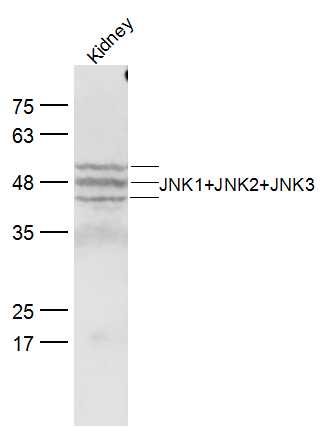 Sample:
Kidney (Mouse) Lysate at 40 ug
Primary: Anti-JNK1+JNK2+JNK3 (SL2592R) at 1/300 dilution
Secondary: IRDye800CW Goat Anti-Rabbit IgG at 1/20000 dilution
Predicted band size: 42-47 kD
Observed band size:42-52 kD
 Sample:
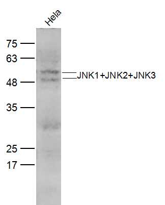
Hela(Human) CellLysate at 30 ug
Primary: Anti-JNK1+JNK2+JNK3 (SL2592R) at 1/300 dilution
Secondary: IRDye800CW Goat Anti-Rabbit IgG at 1/20000 dilution
Predicted band size: 42-47 kD
Observed band size:42-52 kD
 Sample:
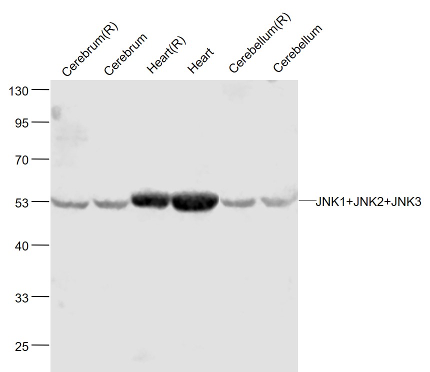
Cerebrum(Rat) Lysate at 40 ug
Cerebrum(Mouse) Lysate at 40 ug
Heart(Rat) Lysate at 40 ug
Heart(Mouse) Lysate at 40 ug
Cerebellum(Rat) Lysate at 40 ug
Cerebellum(Mouse) Lysate at 40 ug
Primary: Anti-JNK1+JNK2+JNK3 (SL2592R) at 1/1000 dilution
Secondary: IRDye800CW Goat Anti-Rabbit IgG at 1/20000 dilution
Predicted band size: 46'54 kD
Observed band size: 54 kD
 Sample:
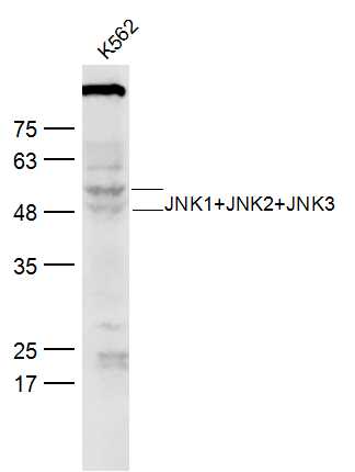
K562 (Human) Lysate at 30 ug
Primary: Anti-JNK1+JNK2+JNK3 (SL2592R) at 1/300 dilution
Secondary: IRDye800CW Goat Anti-Rabbit IgG at 1/20000 dilution
Predicted band size: 42-47 kD
Observed band size:42-52 kD
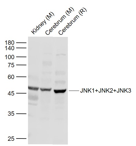 Sample:
Lane 1: Kidney (Mouse) Lysate at 40 ug
Lane 2: Cerebrum (Mouse) Lysate at 40 ug
Lane 3: Cerebrum (Rat) Lysate at 40 ug
Primary: Anti-JNK1+JNK2+JNK3 (SL2592R) at 1/1000 dilution
Secondary: IRDye800CW Goat Anti-Rabbit IgG at 1/20000 dilution
Predicted band size: 45/54 kD
Observed band size: 48 kD
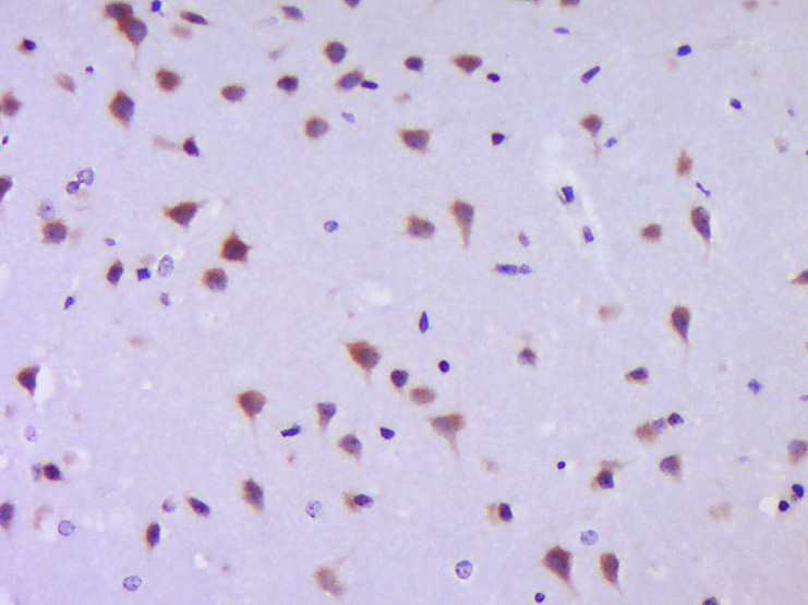
 Paraformaldehyde-fixed, paraffin embedded (mouse brain); Antigen retrieval by boiling in sodium citrate buffer (pH6.0) for 15min; Block endogenous peroxidase by 3% hydrogen peroxide for 20 minutes; Blocking buffer (normal goat serum) at 37°C for 30min; Antibody incubation with (JNK1+JNK2+JNK3) Polyclonal Antibody, Unconjugated (SL2592R) at 1:200 overnight at 4°C, followed by operating according to SP Kit(Rabbit) (sp-0023) instructionsand DAB staining.
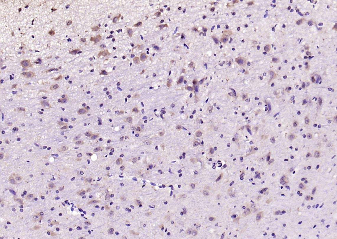 Paraformaldehyde-fixed, paraffin embedded (rat brain); Antigen retrieval by boiling in sodium citrate buffer (pH6.0) for 15min; Block endogenous peroxidase by 3% hydrogen peroxide for 20 minutes; Blocking buffer (normal goat serum) at 37°C for 30min; Antibody incubation with (JNK1+JNK2+JNK3) Polyclonal Antibody, Unconjugated (SL2592R) at 1:200 overnight at 4°C, followed by operating according to SP Kit(Rabbit) (sp-0023) instructionsand DAB staining.
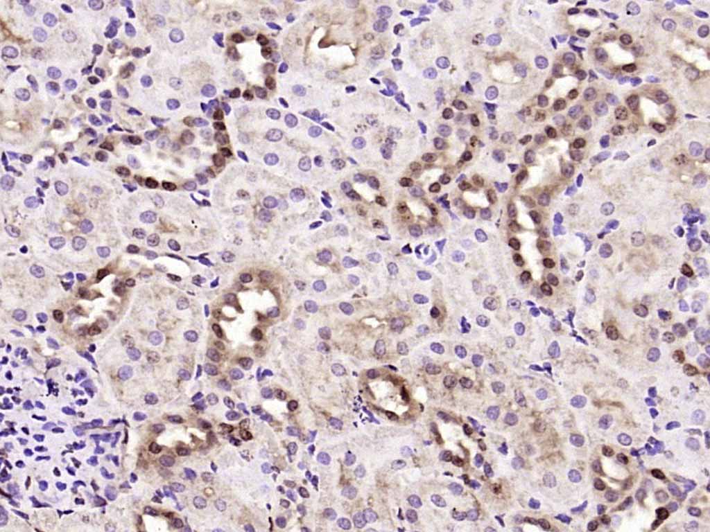 Paraformaldehyde-fixed, paraffin embedded (rat kidney); Antigen retrieval by boiling in sodium citrate buffer (pH6.0) for 15min; Block endogenous peroxidase by 3% hydrogen peroxide for 20 minutes; Blocking buffer (normal goat serum) at 37°C for 30min; Antibody incubation with (JNK1+JNK2+JNK3) Polyclonal Antibody, Unconjugated (SL2592R) at 1:200 overnight at 4°C, followed by operating according to SP Kit(Rabbit) (sp-0023) instructionsand DAB staining.
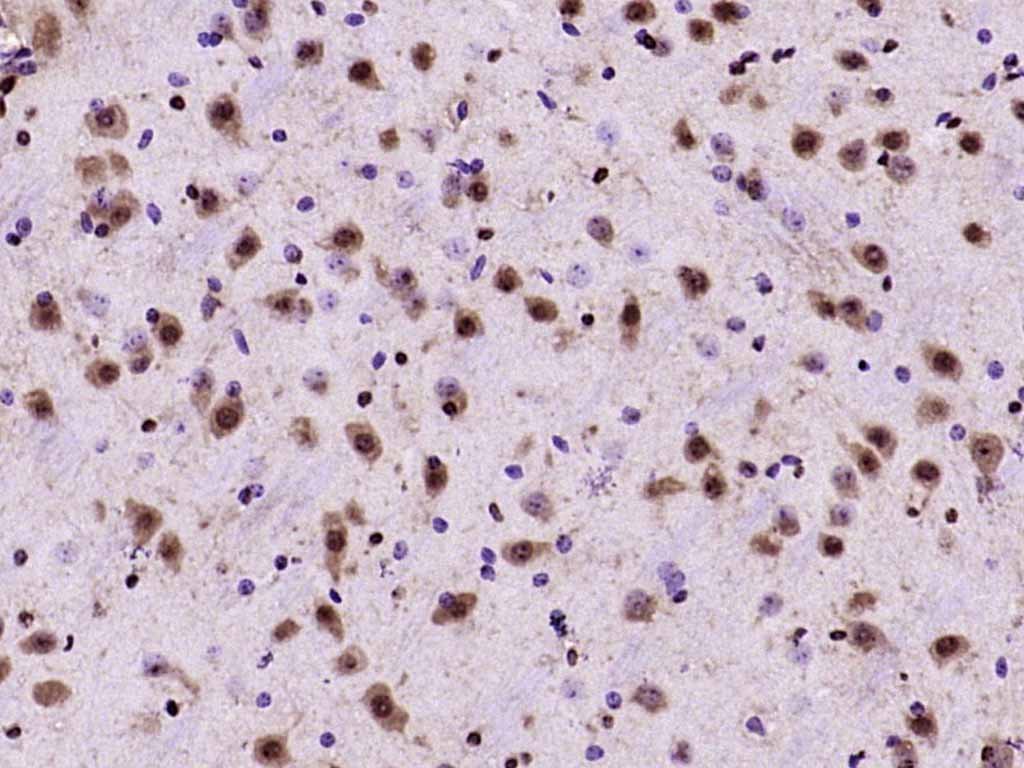 Paraformaldehyde-fixed, paraffin embedded (mouse brain); Antigen retrieval by boiling in sodium citrate buffer (pH6.0) for 15min; Block endogenous peroxidase by 3% hydrogen peroxide for 20 minutes; Blocking buffer (normal goat serum) at 37°C for 30min; Antibody incubation with (JNK1+JNK2+JNK3) Polyclonal Antibody, Unconjugated (SL2592R) at 1:200 overnight at 4°C, followed by operating according to SP Kit(Rabbit) (sp-0023) instructionsand DAB staining.
 Paraformaldehyde-fixed, paraffin embedded (rat cerebellum); Antigen retrieval by boiling in sodium citrate buffer (pH6.0) for 15min; Block endogenous peroxidase by 3% hydrogen peroxide for 20 minutes; Blocking buffer (normal goat serum) at 37°C for 30min; Antibody incubation with (JNK1+JNK2+JNK3) Polyclonal Antibody, Unconjugated (SL2592R) at 1:200 overnight at 4°C, followed by operating according to SP Kit(Rabbit) (sp-0023) instructionsand DAB staining.
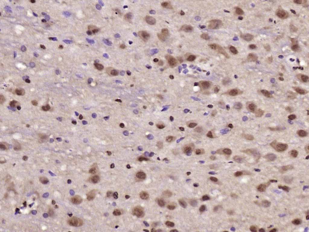 Paraformaldehyde-fixed, paraffin embedded (rat brain); Antigen retrieval by boiling in sodium citrate buffer (pH6.0) for 15min; Block endogenous peroxidase by 3% hydrogen peroxide for 20 minutes; Blocking buffer (normal goat serum) at 37°C for 30min; Antibody incubation with (JNK1+JNK2+JNK3) Polyclonal Antibody, Unconjugated (SL2592R) at 1:200 overnight at 4°C, followed by operating according to SP Kit(Rabbit) (sp-0023) instructionsand DAB staining.
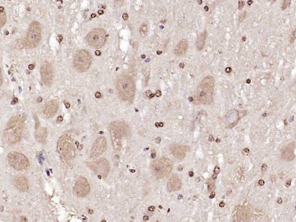 Paraformaldehyde-fixed, paraffin embedded (mouse cerebellum); Antigen retrieval by boiling in sodium citrate buffer (pH6.0) for 15min; Block endogenous peroxidase by 3% hydrogen peroxide for 20 minutes; Blocking buffer (normal goat serum) at 37°C for 30min; Antibody incubation with (JNK1+JNK2+JNK3) Polyclonal Antibody, Unconjugated (SL2592R) at 1:200 overnight at 4°C, followed by operating according to SP Kit(Rabbit) (sp-0023) instructionsand DAB staining.
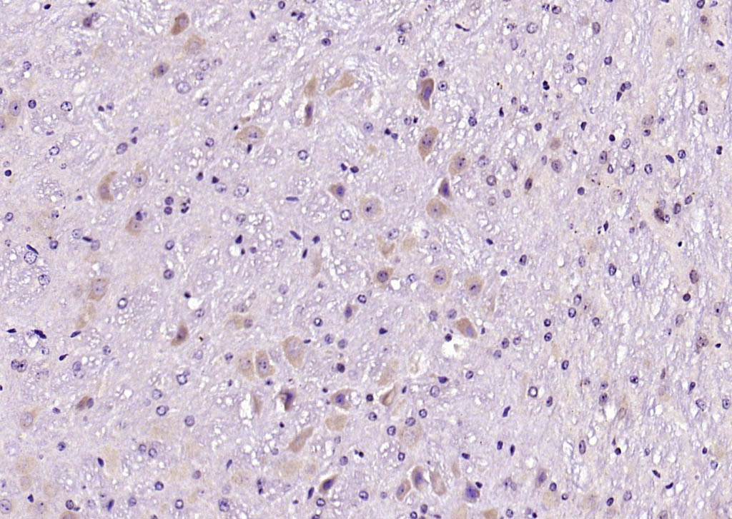
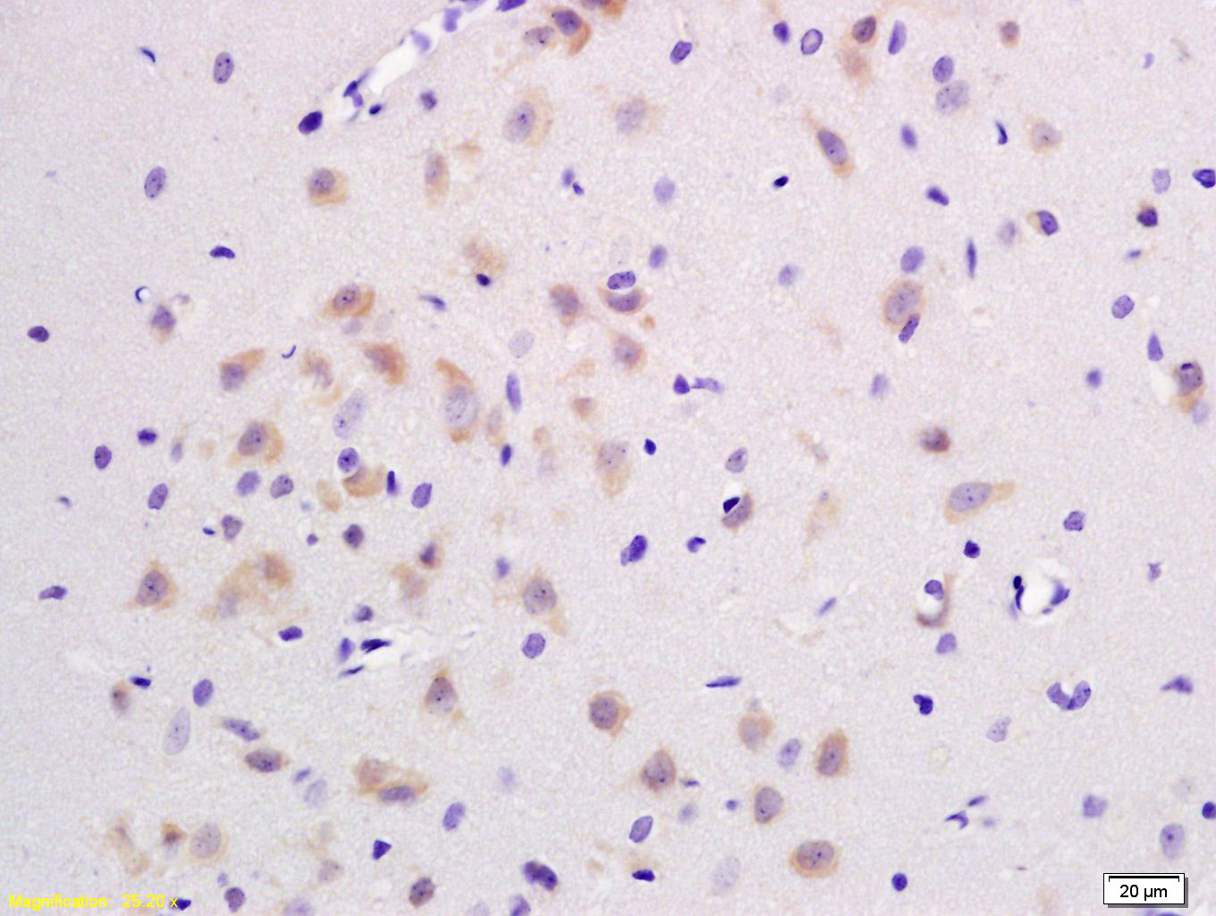 Tissue/cell: rat brain tissue; 4% Paraformaldehyde-fixed and paraffin-embedded;
Antigen retrieval: citrate buffer ( 0.01M, pH 6.0 ), Boiling bathing for 15min; Block endogenous peroxidase by 3% Hydrogen peroxide for 30min; Blocking buffer (normal goat serum,C-0005) at 37℃ for 20 min;
Incubation: Anti-JNK1/2/3 Polyclonal Antibody, Unconjugated(SL2592R) 1:200, overnight at 4°C, followed by conjugation to the secondary antibody(SP-0023) and DAB(C-0010) staining
 Paraformaldehyde-fixed, paraffin embedded (mouse brain tissue); Antigen retrieval by boiling in sodium citrate buffer (pH6.0) for 15min; Block endogenous peroxidase by 3% hydrogen peroxide for 20 minutes; Blocking buffer (normal goat serum) at 37°C for 30min; Antibody incubation with (JNK1+JNK2+JNK3) Polyclonal Antibody, Unconjugated (SL2592R) at 1:400 overnight at 4°C, followed by a conjugated secondary (sp-0023) for 20 minutes and DAB staining.
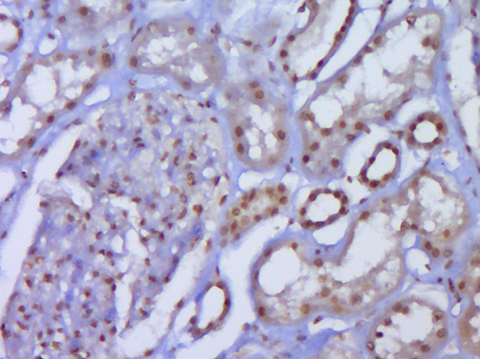 Paraformaldehyde-fixed, paraffin embedded (Human kidney); Antigen retrieval by boiling in sodium citrate buffer (pH6.0) for 15min; Block endogenous peroxidase by 3% hydrogen peroxide for 20 minutes; Blocking buffer (normal goat serum) at 37°C for 30min; Antibody incubation with (JNK1+JNK2+JNK3) Polyclonal Antibody, Unconjugated (SL2592R) at 1:400 overnight at 4°C, followed by operating according to SP Kit(Rabbit) (sp-0023) instructionsand DAB staining.
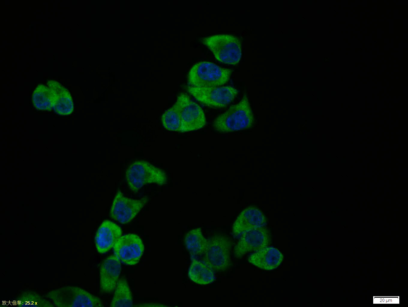 Hela cell; 4% Paraformaldehyde-fixed; Triton X-100 at room temperature for 20 min; Blocking buffer (normal goat serum, C-0005) at 37°C for 20 min; Antibody incubation with (JNK1+JNK2+JNK3) polyclonal Antibody, Unconjugated (SL2592R) 1:100, 90 minutes at 37°C; followed by a conjugated Goat Anti-Rabbit IgG antibody at 37°C for 90 minutes, DAPI (blue, C02-04002) was used to stain the cell nuclei.
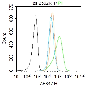 Blank control: Jurkat.
Primary Antibody (green line): Rabbit Anti-JNK1+JNK2+JNK3 antibody (SL2592R)
Dilution: 1μg /10^6 cells;
Isotype Control Antibody (orange line): Rabbit IgG .
Secondary Antibody : Goat anti-rabbit IgG-AF647
Dilution: 1μg /test.
Protocol
The cells were fixed with 4% PFA (10min at room temperature)and then permeabilized with 90% ice-cold methanol for 20 min at-20℃. The cells were then incubated in 5%BSA to block non-specific protein-protein interactions for 30 min at room temperature .Cells stained with Primary Antibody for 30 min at room temperature. The secondary antibody used for 40 min at room temperature. Acquisition of 20,000 events was performed.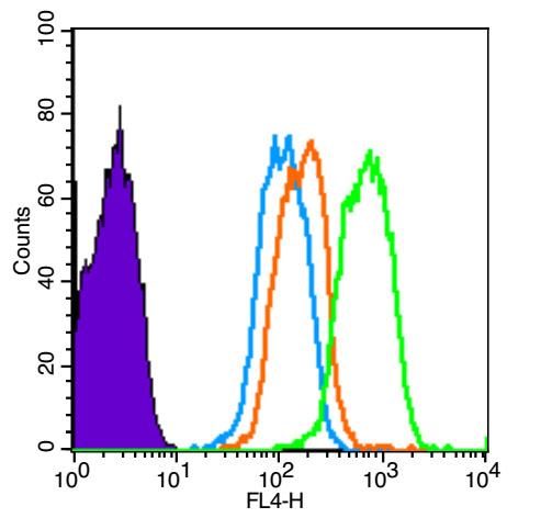 Blank control (Black line): HUVEC (Black). Primary Antibody (green line): Rabbit Anti-JNK1+JNK2+JNK3 antibody (SL2592R) Dilution: 3μg /10^6 cells; Isotype Control Antibody (orange line): Rabbit IgG . Secondary Antibody (white blue line): Goat anti-rabbit IgG-AF647 Dilution: 1μg /test. Protocol The cells were fixed with 4% PFA (10min at room temperature)and then permeabilized with 90% ice-cold methanol for 20 min at room temperature. The cells were then incubated in 5%BSA to block non-specific protein-protein interactions for 30 min at room temperature .Cells stained with Primary Antibody for 30 min at room temperature. The secondary antibody used for 40 min at room temperature. Acquisition of 20,000 events was performed.
Cartpieces
Totalgoods,subtotals:¥Checkout
References (0)
No References
Bought notes(bought amounts latest0)
No one bought this product
User Comment(Total0User Comment Num)
- No comment


 +86 571 56623320
 +86 18668110335

