
Rabbit Anti-Fbx32 antibody
Fbx32; 4833442G10Rik; AI430017; Atrogin 1; Atrogin-1; ATROGIN1; Atrophy gene 1; F box only protein 32; F-box only protein 32; F-box protein 32; FBX32_HUMAN; fbxo25; FBXO32; FLJ32424; MAFbx; MGC108443; MGC137646; MGC33610; Muscle atrophy F box; Muscle atro
View History [Clear]
Details
Product Name Fbx32 Chinese Name Ubiquitin蛋白连接酶抗体 Alias Fbx32; 4833442G10Rik; AI430017; Atrogin 1; Atrogin-1; ATROGIN1; Atrophy gene 1; F box only protein 32; F-box only protein 32; F-box protein 32; FBX32_HUMAN; fbxo25; FBXO32; FLJ32424; MAFbx; MGC108443; MGC137646; MGC33610; Muscle atrophy F box; Muscle atrophy F box protein; Muscle atrophy F-box protein. literatures Research Area Cell biology Stem cells Ubiquitin Immunogen Species Rabbit Clonality Polyclonal React Species Human, Rat, (predicted: Mouse, Dog, Pig, Cow, Horse, Rabbit, ) Applications ELISA=1:5000-10000 IHC-P=1:100-500 IHC-F=1:100-500 Flow-Cyt=0.2ug/test IF=1:100-500 (Paraffin sections need antigen repair)
not yet tested in other applications.
optimal dilutions/concentrations should be determined by the end user.Theoretical molecular weight 42kDa Cellular localization The nucleus cytoplasmic Form Liquid Concentration 1mg/ml immunogen KLH conjugated synthetic peptide derived from human MAFbx: 31-130/355 Lsotype IgG Purification affinity purified by Protein A Buffer Solution 0.01M TBS(pH7.4) with 1% BSA, 0.03% Proclin300 and 50% Glycerol. Storage Shipped at 4℃. Store at -20 °C for one year. Avoid repeated freeze/thaw cycles. Attention This product as supplied is intended for research use only, not for use in human, therapeutic or diagnostic applications. PubMed PubMed Product Detail Fbx32 is an E3 ubiquitin ligase that initiates ATP dependent ubiquitin-mediated proteolysis and promotes muscle atrophy. It is highly expressed during muscle atrophy, whereas mice deficient in this gene were found to be resistant to atrophy. It is also thought to recognize and bind to some phosphorylated proteins and promote their ubiquitination and degradation during skeletal muscle atrophy. Fbx32 interacts with MyoD by ubiquitination via a sequence found in transcriptional coactivators and therefore may play an important role in the course of muscle differentiation by determining the abundance of MyoD.
Function:
Substrate recognition component of a (SKP1-CUL1-F-box protein) E3 ubiquitin-protein ligase complex which mediates the ubiquitination and subsequent proteasomal degradation of target proteins. Probably recognizes and binds to phosphorylated target proteins during skeletal muscle atrophy. Recognizes TERF1.
Subunit:
Part of the SCF (SKP1-CUL1-F-box) E3 ubiquitin-protein ligase complex SCF(FBXO32) formed of CUL1, SKP1, RBX1 and FBXO32.
Subcellular Location:
Cytoplasm. Nucleus. Note=Shuttles between Cytoplasm and the nucleus.
Tissue Specificity:
Specifically expressed in cardiac and skeletal muscle.
Similarity:
Contains 1 F-box domain.
SWISS:
Q969P5
Gene ID:
114907
Database links:
Entrez Gene: 114907 Human
Entrez Gene: 67731 Mouse
Omim: 606604 Human
SwissProt: Q969P5 Human
SwissProt: Q9CPU7 Mouse
Unigene: 403933 Human
Unigene: 292042 Mouse
Unigene: 72619 Rat
Product Picture 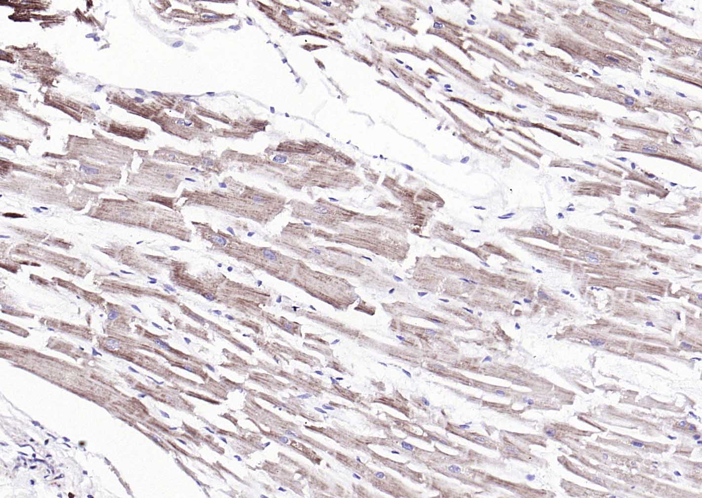 Paraformaldehyde-fixed, paraffin embedded (human heart); Antigen retrieval by boiling in sodium citrate buffer (pH6.0) for 15min; Block endogenous peroxidase by 3% hydrogen peroxide for 20 minutes; Blocking buffer (normal goat serum) at 37°C for 30min; Antibody incubation with (Fbx32) Polyclonal Antibody, Unconjugated (SL2591R) at 1:200 overnight at 4°C, followed by operating according to SP Kit(Rabbit) (sp-0023) instructionsand DAB staining.
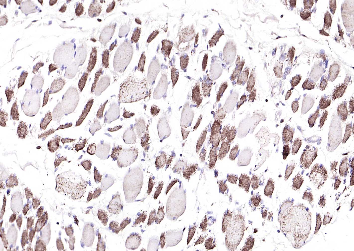 Paraformaldehyde-fixed, paraffin embedded (human skeletal muscle); Antigen retrieval by boiling in sodium citrate buffer (pH6.0) for 15min; Block endogenous peroxidase by 3% hydrogen peroxide for 20 minutes; Blocking buffer (normal goat serum) at 37°C for 30min; Antibody incubation with (Fbx32) Polyclonal Antibody, Unconjugated (SL2591R) at 1:200 overnight at 4°C, followed by operating according to SP Kit(Rabbit) (sp-0023) instructionsand DAB staining.
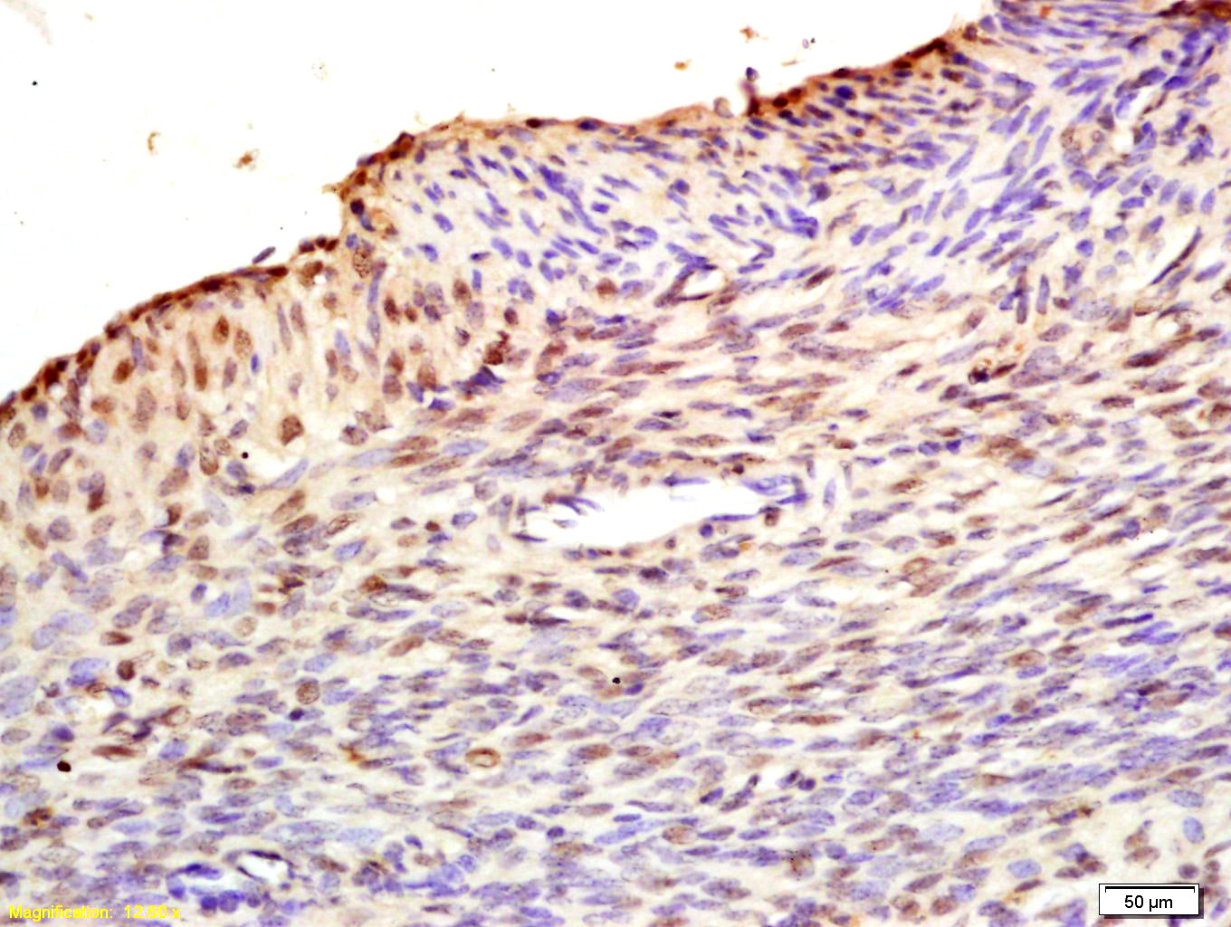 Tissue/cell: rat uterus tissue; 4% Paraformaldehyde-fixed and paraffin-embedded;
Antigen retrieval: citrate buffer ( 0.01M, pH 6.0 ), Boiling bathing for 15min; Block endogenous peroxidase by 3% Hydrogen peroxide for 30min; Blocking buffer (normal goat serum,C-0005) at 37℃ for 20 min;
Incubation: Anti-MAFbx/Fbx32 Polyclonal Antibody, Unconjugated(SL2591R) 1:200, overnight at 4°C, followed by conjugation to the secondary antibody(SP-0023) and DAB(C-0010) staining
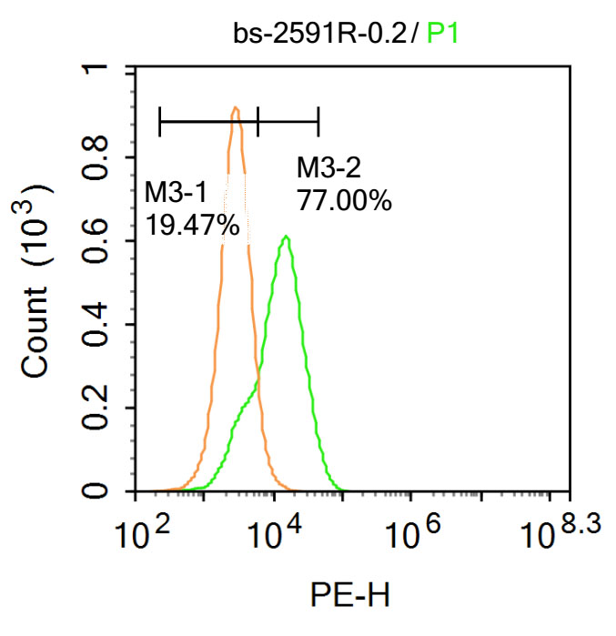 Blank control: Hela.
Primary Antibody (green line): Rabbit Anti-Fbx32 antibody (SL2591R)
Dilution: 1μg /10^6 cells;
Isotype Control Antibody (orange line): Rabbit IgG .
Secondary Antibody : Goat anti-rabbit IgG-PE
Dilution: 1μg /test.
Protocol
The cells were fixed with 4% PFA (10min at room temperature)and then permeabilized with 90% ice-cold methanol for 20 min at-20℃. The cells were then incubated in 5%BSA to block non-specific protein-protein interactions for 30 min at at room temperature .Cells stained with Primary Antibody for 30 min at room temperature. The secondary antibody used for 40 min at room temperature. Acquisition of 20,000 events was performed.
Cartpieces
Totalgoods,subtotals:¥Checkout
References (0)
No References
Bought notes(bought amounts latest0)
No one bought this product
User Comment(Total0User Comment Num)
- No comment


 +86 571 56623320
 +86 18668110335

