
Rabbit Anti-CD26 antibody
ADCP2; DPPIV; Dipeptidyl peptidase 4; ADABP; ADCP 2; Adenosine deaminase complexing protein 2; CD 26; CD26 antigen 3; Dipeptidyl peptidase 4; Dipeptidyl peptidase iv; Dipeptidylpeptidase 4; Dipeptidylpeptidase IV; DPP 4; DPP IV; DPP4; Intestinal dipeptidy
View History [Clear]
Details
Product Name CD26 Chinese Name CD26/DPP4抗体 Alias ADCP2; DPPIV; Dipeptidyl peptidase 4; ADABP; ADCP 2; Adenosine deaminase complexing protein 2; CD 26; CD26 antigen 3; Dipeptidyl peptidase 4; Dipeptidyl peptidase iv; Dipeptidylpeptidase 4; Dipeptidylpeptidase IV; DPP 4; DPP IV; DPP4; Intestinal dipeptidyl peptidase; T cell activation antigen CD26; TP 103; TP103; DPP4_HUMAN. literatures Research Area Tumour Cell biology immunology Stem cells Apoptosis The cell membrane受体 Cell adhesion molecule Cell Surface Molecule glycoprotein lymphocyte t-lymphocyte b-lymphocyte Immunogen Species Rabbit Clonality Polyclonal React Species Human, Rat, (predicted: Mouse, Pig, Cow, Horse, Rabbit, ) Applications ELISA=1:5000-10000 IHC-P=1:100-500 Flow-Cyt=3μg/Test (Paraffin sections need antigen repair)
not yet tested in other applications.
optimal dilutions/concentrations should be determined by the end user.Theoretical molecular weight 84kDa Detection molecular weight 110 kDa Cellular localization cytoplasmic The cell membrane Extracellular matrix Secretory protein Form Liquid Concentration 1mg/ml immunogen KLH conjugated synthetic peptide derived from human CD26: 661-766/766 Lsotype IgG Purification affinity purified by Protein A Buffer Solution 0.01M TBS(pH7.4) with 1% BSA, 0.03% Proclin300 and 50% Glycerol. Storage Shipped at 4℃. Store at -20 °C for one year. Avoid repeated freeze/thaw cycles. Attention This product as supplied is intended for research use only, not for use in human, therapeutic or diagnostic applications. PubMed PubMed Product Detail The DPP4 gene encodes dipeptidyl peptidase 4, which is identical to adenosine deaminase complexing protein-2, and to the T-cell activation antigen CD26. It is an intrinsic type II transmembrane glycoprotein and a serine exopeptidase that cleaves X-proline dipeptides from the N-terminus of polypeptides. Dipeptidyl peptidase 4 is highly involved in glucose and insulin metabolism, as well as in immune regulation. This protein was shown to be a functional receptor for Middle East respiratory syndrome coronavirus (MERS-CoV), and protein modeling suggests that it may play a similar role with SARS-CoV-2, the virus responsible for COVID-19. [provided by RefSeq, Apr 2020]
Function:
Cell surface glycoprotein receptor involved in the costimulatory signal essential for T-cell receptor (TCR)-mediated T-cell activation. Acts as a positive regulator of T-cell coactivation, by binding at least ADA, CAV1, IGF2R, and PTPRC. Its binding to CAV1 and CARD11 induces T-cell proliferation and NF-kappa-B activation in a T-cell receptor/CD3-dependent manner. Its interaction with ADA also regulates lymphocyte-epithelial cell adhesion. In association with FAP is involved in the pericellular proteolysis of the extracellular matrix (ECM), the migration and invasion of endothelial cells into the ECM. May be involved in the promotion of lymphatic endothelial cells adhesion, migration and tube formation. When overexpressed, enhanced cell proliferation, a process inhibited by GPC3. Acts also as a serine exopeptidase with a dipeptidyl peptidase activity that regulates various physiological processes by cleaving peptides in the circulation, including many chemokines, mitogenic growth factors, neuropeptides and peptide hormones. Removes N-terminal dipeptides sequentially from polypeptides having unsubstituted N-termini provided that the penultimate residue is proline.
Subcellular Location:
Cell membrane. Apical cell membrane. Cell projection > invadopodium membrane. Cell projection > lamellipodium membrane. Cell junction. Membrane raft. Translocated to the apical membrane through the concerted action of N- and O-Glycans and its association with lipid microdomains containing cholesterol and sphingolipids. Redistributed to membrane rafts in T-cell in a interleukin-12-dependent activation. Its interaction with CAV1 is necessary for its translocation to membrane rafts. Colocalized with PTPRC in membrane rafts. Colocalized with FAP in invadopodia and lamellipodia of migratory activated endothelial cells in collagenous matrix. Colocalized with FAP on endothelial cells of capillary-like microvessels but not large vessels within invasive breast ductal carcinoma. Colocalized with ADA at the cell junction in lymphocyte-epithelial cell adhesion. Colocalized with IGF2R in internalized cytoplasmic vesicles adjacent to the cell surface and Secreted. Detected in the serum and the seminal fluid.
Tissue Specificity:
Expressed specifically in lymphatic vessels but not in blood vessels in the skin, small intestine, esophagus, ovary, breast and prostate glands. Not detected in lymphatic vessels in the lung, kidney, uterus, liver and stomach (at protein level). Expressed in the poorly differentiated crypt cells of the small intestine as well as in the mature villous cells. Expressed at very low levels in the colon.
Post-translational modifications:
The soluble form (Dipeptidyl peptidase 4 soluble form also named SDPP) derives from the membrane form (Dipeptidyl peptidase 4 membrane form also named MDPP) by proteolytic processing.
N- and O-Glycosylated.
Phosphorylated. Mannose 6-phosphate residues in the carbohydrate moiety are necessary for interaction with IGF2R in activated T-cells. Mannose 6-phosphorylation is induced during T-cell activation.
Similarity:
Belongs to the peptidase S9B family. DPPIV subfamily.
SWISS:
P27487
Gene ID:
1803
Database links:Entrez Gene: 1803 Human
Entrez Gene: 13482 Mouse
Omim: 102720 Human
SwissProt: P27487 Human
SwissProt: P28843 Mouse
Unigene: 368912 Human
Unigene: 1151 Mouse
Unigene: 91364 Rat
CD26是细胞表面上一种氨基肽酶,具有丝氨酸蛋白酶活性,属于丝氨酸蛋白酶s9b家族,分布于多种细胞表面或游离于胞浆中,具有高度保守的蛋白水解酶活性. 还是一种The cell membrane受体、共刺激分子,参与机体免疫调节、细胞迁移、细胞黏附和Apoptosis.Product Picture 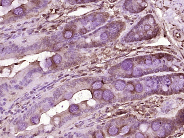 Paraformaldehyde-fixed, paraffin embedded (rat intestine tissue); Antigen retrieval by boiling in sodium citrate buffer (pH6.0) for 15min; Block endogenous peroxidase by 3% hydrogen peroxide for 20 minutes; Blocking buffer (normal goat serum) at 37°C for 30min; Antibody incubation with (CD26) Polyclonal Antibody, Unconjugated (SL2570R) at 1:400 overnight at 4°C, followed by operating according to SP Kit(Rabbit) (sp-0023) instructionsand DAB staining.
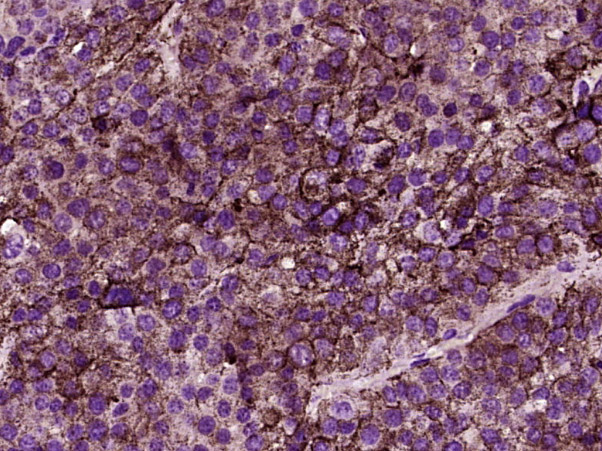 Paraformaldehyde-fixed, paraffin embedded (Human melanoma); Antigen retrieval by boiling in sodium citrate buffer (pH6.0) for 15min; Block endogenous peroxidase by 3% hydrogen peroxide for 20 minutes; Blocking buffer (normal goat serum) at 37°C for 30min; Antibody incubation with (CD26) Polyclonal Antibody, Unconjugated (SL2570R) at 1:400 overnight at 4°C, followed by operating according to SP Kit(Rabbit) (sp-0023) instructionsand DAB staining.
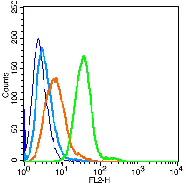 Blank control(blue): Raji (fixed with 2% paraformaldehyde (10 min)).
Primary Antibody:Rabbit Anti-CD26 antibody(SL2570R), Dilution: 3μg in 100 μL 1X PBS containing 0.5% BSA;
Isotype Control Antibody: Rabbit IgG(orange) ,used under the same conditions );
Secondary Antibody: Goat anti-rabbit IgG-PE(white blue), Dilution: 1:200 in 1 X PBS containing 0.5% BSA.
Cartpieces
Totalgoods,subtotals:¥Checkout
References (0)
No References
Bought notes(bought amounts latest0)
No one bought this product
User Comment(Total0User Comment Num)
- No comment


 +86 571 56623320
 +86 18668110335

