
Rabbit Anti-DEP1 antibody
CD 148; CD148; CD148 antigen; Density enhanced phosphatase 1; Density-enhanced phosphatase 1; DEP 1; DEP-1; HPTP eta; HPTPeta; Human density enhanced phosphatase 1; Protein tyrosine phosphatase eta; Protein tyrosine phosphatase receptor type J; Protein ty
View History [Clear]
Details
Product Name DEP1 Chinese Name 蛋白酪氨酸磷酸酶CD148抗体 Alias CD 148; CD148; CD148 antigen; Density enhanced phosphatase 1; Density-enhanced phosphatase 1; DEP 1; DEP-1; HPTP eta; HPTPeta; Human density enhanced phosphatase 1; Protein tyrosine phosphatase eta; Protein tyrosine phosphatase receptor type J; Protein tyrosine phosphatase receptor type J polypeptide; Protein-tyrosine phosphatase eta; Protein-tyrosine phosphatase receptor type J; PTPJ; Ptprj; PTPRJ_HUMAN; R PTP Eta; R-PTP-eta; R-PTP-J; Receptor type tyrosine protein phosphatase eta; Receptor-type tyrosine-protein phosphatase eta; SCC 1; SCC1. Research Area Cell biology Signal transduction Kinases and Phosphatases Immunogen Species Rabbit Clonality Polyclonal React Species Human, Mouse, (predicted: Rat, Rabbit, ) Applications WB=1:500-2000 ELISA=1:5000-10000 IHC-P=1:100-500 (Paraffin sections need antigen repair)
not yet tested in other applications.
optimal dilutions/concentrations should be determined by the end user.Theoretical molecular weight 147kDa Cellular localization The cell membrane Form Liquid Concentration 1mg/ml immunogen KLH conjugated synthetic peptide derived from human PTPRJ: 865-970/1337 <Extracellular> Lsotype IgG Purification affinity purified by Protein A Buffer Solution 0.01M TBS(pH7.4) with 1% BSA, 0.03% Proclin300 and 50% Glycerol. Storage Shipped at 4℃. Store at -20 °C for one year. Avoid repeated freeze/thaw cycles. Attention This product as supplied is intended for research use only, not for use in human, therapeutic or diagnostic applications. PubMed PubMed Product Detail The protein encoded by this gene is a member of the protein tyrosine phosphatase (PTP) family. PTPs are known to be signaling molecules that regulate a variety of cellular processes, including cell growth, differentiation, mitotic cycle, and oncogenic transformation. This PTP possesses an extracellular region containing five fibronectin type III repeats, a single transmembrane region, and a single intracytoplasmic catalytic domain, and thus represents a receptor-type PTP. This protein is present in all hematopoietic lineages, and was shown to negatively regulate T cell receptor signaling possibly through interfering with the phosphorylation of Phospholipase C Gamma 1 and Linker for Activation of T Cells. This protein can also dephosphorylate the PDGF beta receptor, and may be involved in UV-induced signal transduction. Multiple transcript variants encoding different isoforms have been found for this gene. [provided by RefSeq].
Function:
Tyrosine phosphatase which dephosphorylates or contributes to the dephosphorylation of CTNND1, FLT3, PDGFRB, MET, RET (variant MEN2A), KDR, LYN, SRC, MAPK1, MAPK3, EGFR, TJP1, OCLN, PIK3R1 and PIK3R2. Plays a role in cell adhesion, migration, proliferation and differentiation. Involved in vascular development. Regulator of macrophage adhesion and spreading. Positively affects cell-matrix adhesion. Positive regulator of platelet activation and thrombosis. Negative regulator of cell proliferation. Negative regulator of PDGF-stimulated cell migration; through dephosphorylation of PDGFR. Positive regulator of endothelial cell survival, as well as of VEGF-induced SRC and AKT activation; through KDR dephosphorylation. Negative regulator of EGFR signaling pathway; through EGFR dephosphorylation. Enhances the barrier function of epithelial junctions during reassembly. Negatively regulates T-cell receptor (TCR) signaling. Upon T-cell TCR activation, it is up-regulated and excluded from the immunological synapses, while upon T-cell-antigen presenting cells (APC) disengagement, it is no longer excluded and can dephosphorylate PLCG1 and LAT to down-regulate prolongation of signaling.
Subunit:
Monomer. Interacts with CTNNB1 (phosphorylated) and JUP (phosphorylated). Interacts with FLT3 (phosphorylated). Interacts with GAB1 and GRB2.
Subcellular Location:
Cell membrane.
Tissue Specificity:
Expressed in the promyelocytic cell line HL60, the granulocyte-macrophage colony-stimulating factor-dependent leukemic cell line F-36P, and the interleukin-3 and erythropoietin-dependent leukemic cell line F-36E. Expressed predominantly in epithelial cells and lymphocytes. Enhanced expression at high cell density.
Post-translational modifications:
N- and O-glycosylated.
Similarity:
Belongs to the protein-tyrosine phosphatase family. Receptor class 3 subfamily.
Contains 9 fibronectin type-III domains.
Contains 1 tyrosine-protein phosphatase domain.
SWISS:
Q12913
Gene ID:
5795
Database links:Entrez Gene: 5795 Human
Omim: 600925 Human
SwissProt: Q12913 Human
Unigene: 318547 Human
Product Picture 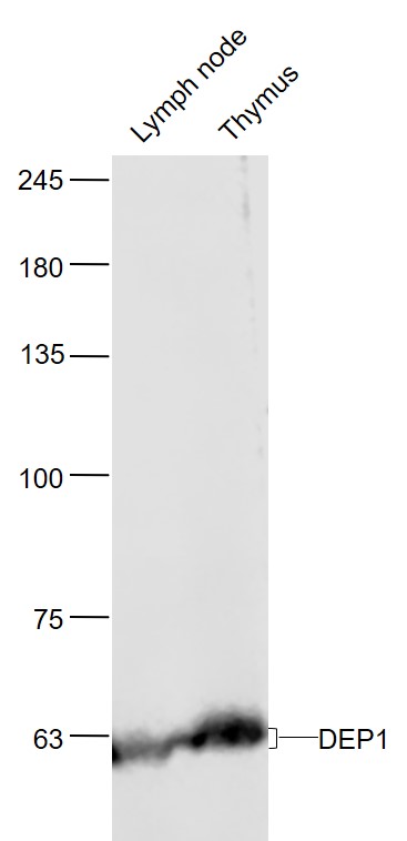 Sample:
Lymph node (Mouse) Lysate at 40 ug
Thymus (Mouse) Lysate at 40 ug
Primary: Anti-DEP1 (SL2567R) at 1/1000 dilution
Secondary: IRDye800CW Goat Anti-Rabbit IgG at 1/20000 dilution
Predicted band size: 60'100 kD
Observed band size: 60 kD
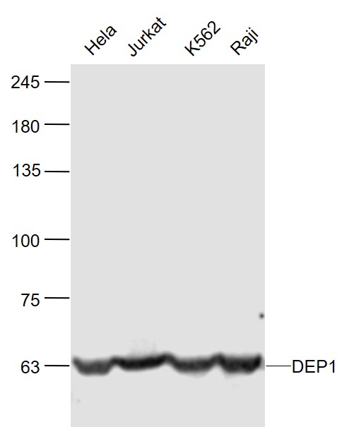 Sample:
Hela(Human) Cell Lysate at 30 ug
Jurkat(Human) Cell Lysate at 30 ug
K562(Human) Cell Lysate at 30 ug
Raji(Human) Cell Lysate at 30 ug
Primary: Anti-DEP1 (SL2567R) at 1/1000 dilution
Secondary: IRDye800CW Goat Anti-Rabbit IgG at 1/20000 dilution
Predicted band size: 60'100 kD
Observed band size: 60 kD
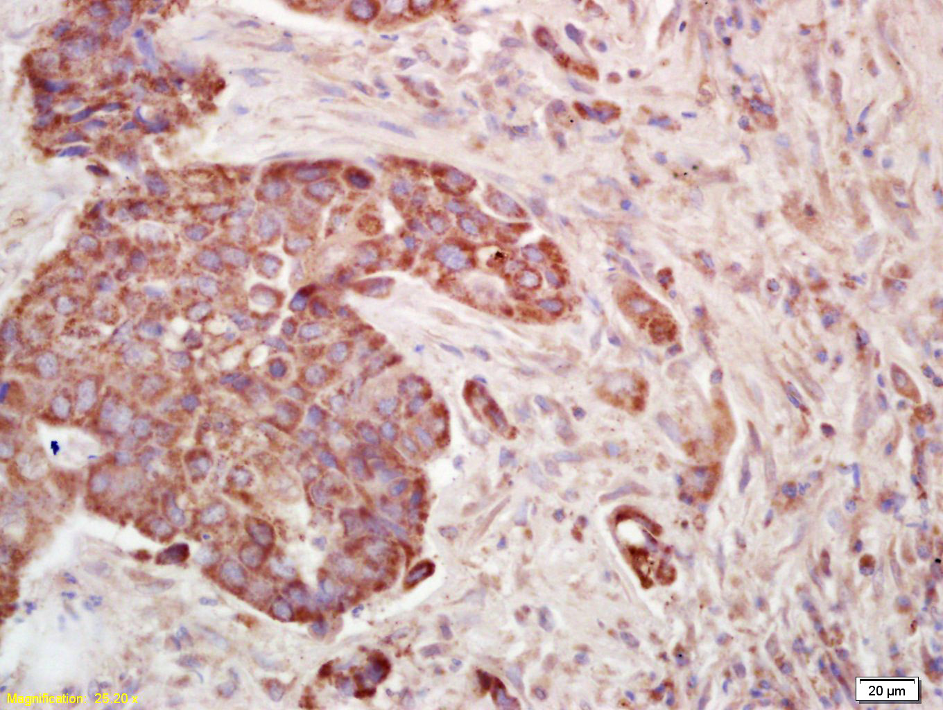 Tissue/cell: human lung carcinoma; 4% Paraformaldehyde-fixed and paraffin-embedded;
Antigen retrieval: citrate buffer ( 0.01M, pH 6.0 ), Boiling bathing for 15min; Block endogenous peroxidase by 3% Hydrogen peroxide for 30min; Blocking buffer (normal goat serum,C-0005) at 37℃ for 20 min;
Incubation: Anti-PTP/PTPase/CD148 Polyclonal Antibody, Unconjugated(SL2567R) 1:200, overnight at 4°C, followed by conjugation to the secondary antibody(SP-0023) and DAB(C-0010) staining
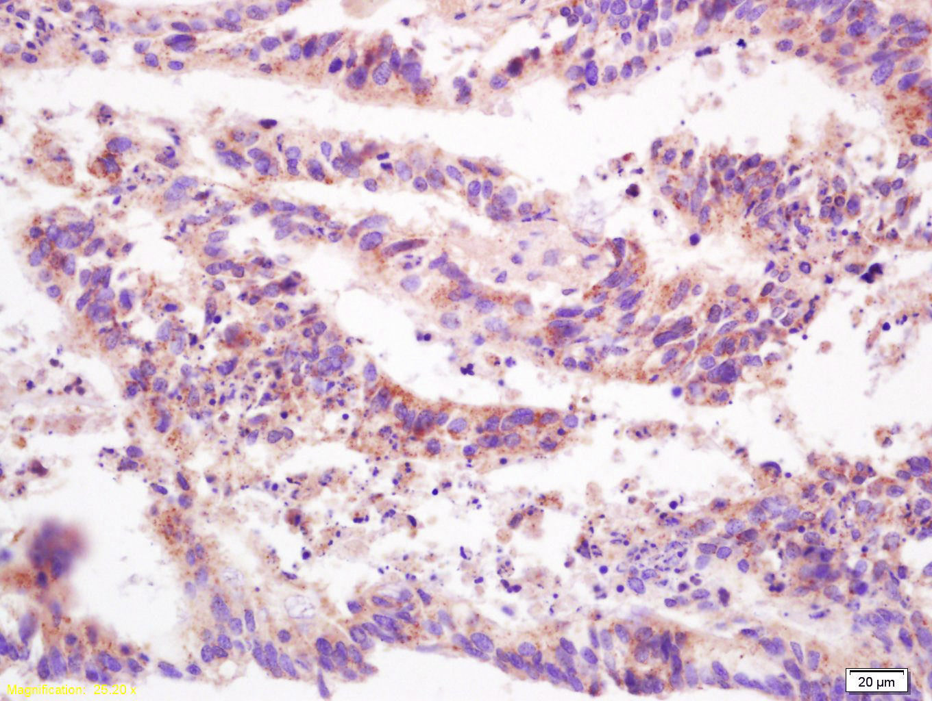 Tissue/cell: human colon carcinoma; 4% Paraformaldehyde-fixed and paraffin-embedded;
Antigen retrieval: citrate buffer ( 0.01M, pH 6.0 ), Boiling bathing for 15min; Block endogenous peroxidase by 3% Hydrogen peroxide for 30min; Blocking buffer (normal goat serum,C-0005) at 37℃ for 20 min;
Incubation: Anti-PTP/PTPase/CD148 Polyclonal Antibody, Unconjugated(SL2567R) 1:200, overnight at 4°C, followed by conjugation to the secondary antibody(SP-0023) and DAB(C-0010) staining
Cartpieces
Totalgoods,subtotals:¥Checkout
References (0)
No References
Bought notes(bought amounts latest0)
No one bought this product
User Comment(Total0User Comment Num)
- No comment


 +86 571 56623320
 +86 18668110335

