
Rabbit Anti-MuRF1 antibody
MTrim63; uRF 1; MuRF-1; Muscle-specific RING finger protein 1; Muscle-specific RING finger protein 1; E3 ubiquitin-protein ligase TRIM63; FLJ32380; IRF; MURF1; MURF 1; MURF2; RNF28; SMRZ; Iris ring finger protein; Muscle specific ring finger protein 2; Ri
View History [Clear]
Details
Product Name MuRF1 Chinese Name 肌肉细胞特异性Ubiquitin蛋白连接酶1抗体 Alias MTrim63; uRF 1; MuRF-1; Muscle-specific RING finger protein 1; Muscle-specific RING finger protein 1; E3 ubiquitin-protein ligase TRIM63; FLJ32380; IRF; MURF1; MURF 1; MURF2; RNF28; SMRZ; Iris ring finger protein; Muscle specific ring finger protein 2; Ring finger protein 28; RNF28; SMRZ; Striated muscle RING zinc finger protein; TRIM 63; TRIM63; Tripartite motif containing 63; Tripartite motif containing protein 63; Ubiquitin ligase TRIM63. literatures Research Area Cardiovascular Cell biology Signal transduction Immunogen Species Rabbit Clonality Polyclonal React Species Human, Mouse, (predicted: Rat, Pig, Cow, Horse, Rabbit, ) Applications ELISA=1:5000-10000 IHC-P=1:100-500 IHC-F=1:100-500 Flow-Cyt=2ug/Test IF=1:100-500 (Paraffin sections need antigen repair)
not yet tested in other applications.
optimal dilutions/concentrations should be determined by the end user.Theoretical molecular weight 39kDa Cellular localization The nucleus cytoplasmic Form Liquid Concentration 1mg/ml immunogen KLH conjugated synthetic peptide derived from human MuRF1: 251-353/353 Lsotype IgG Purification affinity purified by Protein A Buffer Solution 0.01M TBS(pH7.4) with 1% BSA, 0.03% Proclin300 and 50% Glycerol. Storage Shipped at 4℃. Store at -20 °C for one year. Avoid repeated freeze/thaw cycles. Attention This product as supplied is intended for research use only, not for use in human, therapeutic or diagnostic applications. PubMed PubMed Product Detail This gene encodes a member of the RING zinc finger protein family found in striated muscle and iris. The product of this gene is localized to the Z-line and M-line lattices of myofibrils, where titin's N-terminal and C-terminal regions respectively bind to the sarcomere. In vitro binding studies have shown that this protein also binds directly to titin near the region of titin containing kinase activity. Another member of this protein family binds to microtubules. Since these family members can form heterodimers, this suggests that these proteins may serve as a link between titin kinase and microtubule-dependent signal pathways in muscle. [provided by RefSeq].
Function:
E3 ubiquitin ligase. Mediates the ubiquitination and subsequent proteasomal degradation of CKM, GMEB1 and HIBADH. Regulates the proteasomal degradation of muscle proteins under amino acid starvation, where muscle protein is catabolized to provide other organs with amino acids. Inhibits de novo skeletal muscle protein synthesis under amino acid starvation. Regulates proteasomal degradation of cardiac troponin I/TNNI3 and probably of other sarcomeric-associated proteins. May play a role in striated muscle atrophy and hypertrophy by regulating an anti-hypertrophic PKC-mediated signaling pathway. May regulate the organization of myofibrils through TTN in muscle cells.
Subunit:
Homodimer. Homooligomer and heterooligomer. Interacts with SUMO2, titin/TTN and GMEB1. Interacts with TRIM54 and probably with TRIM55 and TNNI3. Forms a ternary complex with GNB2L1 and PRKCE. Interacts with CKM.
Subcellular Location:
Cytoplasm. Nucleus. Cytoplasm, myofibril, sarcomere, M line. Cytoplasm, myofibril, sarcomere, Z line. Note=Colocalizes with TNNI3 in myocytes. Localizes to the M- and Z-lines in skeletal muscle.
Tissue Specificity:
Muscle specific. Selectively expressed in heart and skeletal muscle. Also expressed in the iris.
Similarity:
Contains 1 B box-type zinc finger.
Contains 1 COS domain.
Contains 1 RING-type zinc finger.
SWISS:
Q969Q1
Gene ID:
84676
Database links:Entrez Gene: 84676 Human
Entrez Gene: 433766 Mouse
Omim: 606131 Human
SwissProt: Q969Q1 Human
SwissProt: Q38HM4 Mouse
Unigene: 279709 Human
Unigene: 331961 Mouse
Unigene: 40636 Rat
Product Picture 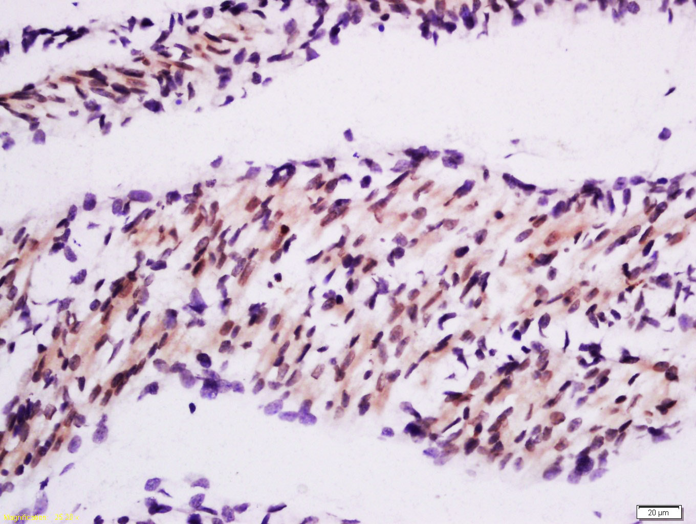 Tissue/cell: mouse embryos tissue; 4% Paraformaldehyde-fixed and paraffin-embedded;
Antigen retrieval: citrate buffer ( 0.01M, pH 6.0 ), Boiling bathing for 15min; Block endogenous peroxidase by 3% Hydrogen peroxide for 30min; Blocking buffer (normal goat serum,C-0005) at 37℃ for 20 min;
Incubation: Anti-MuRF1 Polyclonal Antibody, Unconjugated(SL2539R) 1:200, overnight at 4°C, followed by conjugation to the secondary antibody(SP-0023) and DAB(C-0010) staining
 Blank control:K562.
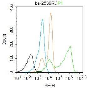
Primary Antibody (green line): Rabbit Anti-MuRF1 antibody (SL2539R)
Dilution: 2μg /10^6 cells;
Isotype Control Antibody (orange line): Rabbit IgG .
Secondary Antibody : Goat anti-rabbit IgG-PE
Dilution: 1μg /test.
Protocol
The cells were fixed with 4% PFA (10min at room temperature)and then permeabilized with 90% ice-cold methanol for 20 min at-20℃. The cells were then incubated in 5%BSA to block non-specific protein-protein interactions for 30 min at room temperature .Cells stained with Primary Antibody for 30 min at room temperature. The secondary antibody used for 40 min at room temperature. Acquisition of 20,000 events was performed.
Cartpieces
Totalgoods,subtotals:¥Checkout
Bought notes(bought amounts latest0)
No one bought this product
User Comment(Total0User Comment Num)
- No comment


 +86 571 56623320
 +86 18668110335

