
Rabbit Anti-CD9 antibody
CD9 antigen; MIC3; MRP-1; BTCC-1; DRAP-27; TSPAN29; TSPAN-29; CD9_HUMAN.
View History [Clear]
Details
Product Name CD9 Chinese Name CD9蛋白抗体 Alias CD9 antigen; MIC3; MRP-1; BTCC-1; DRAP-27; TSPAN29; TSPAN-29; CD9_HUMAN. literatures Research Area Tumour Cell biology immunology Developmental biology Cell Surface Molecule Immunogen Species Rabbit Clonality Polyclonal React Species Human, Mouse, Rat, (predicted: Dog, Pig, Cow, Rabbit, Sheep, ) Applications WB=1:500-2000 ELISA=1:5000-10000 IHC-P=1:100-500 IHC-F=1:100-500 Flow-Cyt=1μg/Test IF=1:100-500 (Paraffin sections need antigen repair)
not yet tested in other applications.
optimal dilutions/concentrations should be determined by the end user.Theoretical molecular weight 24kDa Detection molecular weight 24kDa Cellular localization The cell membrane Form Liquid Concentration 1mg/ml immunogen KLH conjugated synthetic peptide derived from human CD9: 101-200/228 <Extracellular> Lsotype IgG Purification affinity purified by Protein A Buffer Solution 0.01M TBS(pH7.4) with 1% BSA, 0.03% Proclin300 and 50% Glycerol. Storage Shipped at 4℃. Store at -20 °C for one year. Avoid repeated freeze/thaw cycles. Attention This product as supplied is intended for research use only, not for use in human, therapeutic or diagnostic applications. PubMed PubMed Product Detail CD9 antigen is a glycoprotein expressed on the surface of developing B lymphocytes, platelets, monocytes, eosinophils, basophil, stimulated T lymphocytes and by neurons and glial cells in the peripheral nervous system. It belongs to a family of membrane proteins termed tetraspanins which transverse the membrane four times. In pre B cells and platelets, CD9 antigen regulates cell activation and aggregation possibly through an association with the integrin CD41 / CD61 (GPIIb / GPIIIa). It also regulates cell motility in a variety of cell lines, and appears to be an important regulator of Schwann cell behaviour in peripheral nerve.
Function:
Involved in platelet activation and aggregation. Regulates paranodal junction formation. Involved in cell adhesion, cell motility and tumor metastasis. Required for sperm-egg fusion.
Subunit:
Forms both disulfide-linked homodimers and higher homooligomers as well as heterooligomers with other members of the tetraspanin family. Associates with CR2/CD21 and with PTGFRN/CD9P1. Interacts directly with IGSF8.
Subcellular Location:
Membrane; Multi-pass membrane protein.
Tissue Specificity:
Expressed by a variety of hematopoietic and epithelial cells.
Post-translational modifications:
Protein exists in three forms with molecular masses between 22 and 27 kDa, and is known to carry covalently linked fatty acids.
Similarity:
Belongs to the tetraspanin (TM4SF) family.
SWISS:
P21926
Gene ID:
928
Database links:Entrez Gene: 928 Human
Entrez Gene: 12527 Mouse
SwissProt: P21926 Human
SwissProt: P40240 Mouse
CD9是一种普遍存在于The cell membrane表面属于四跨膜超蛋白家族的细胞表面glycoprotein, 参与质膜的融合过程,在The cell membrane生物学中发挥重要作用。
近年来经研究发现,CD9与多种Tumour转移有关,故称Tumour转移抑制基因亦称抗Tumour转移基因。是一个新的Tumour转移抑制基因膜运动相关蛋白-1(CD9/MRP-1),其分子功能还有待于进一步研究。CD9主要在早B细胞、活化Tlymphocyte、嗜酸性粒细胞、嗜碱性粒细胞和血小板表达。Product Picture 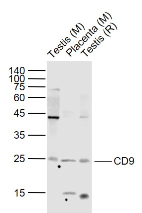 Sample:
Lane 1: Testis (Mouse) Lysate at 40 ug
Lane 2: Placenta (Mouse) Lysate at 40 ug
Lane 3: Testis (Rat) Lysate at 40 ug
Primary:
Anti-CD9 (SL2489R) at 1/1000 dilution
Secondary: IRDye800CW Goat Anti-Rabbit IgG at 1/20000 dilution
Predicted band size: 24 kD
Observed band size: 24 kD
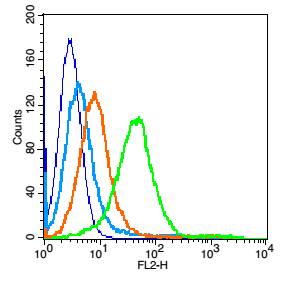 Blank control: Mouse spleen cells(blue).
Primary Antibody:Rabbit Anti-CD9 antibody(SL2489R), Dilution: 1μg in 100 μL 1X PBS containing 0.5% BSA;
Isotype Control Antibody: Rabbit IgG(orange) ,used under the same conditions );
Secondary Antibody: Goat anti-rabbit IgG-PE(white blue), Dilution: 1:200 in 1 X PBS containing 0.5% BSA.
Protocol
Primary antibody were incubated for 30 min on the ice, followed by 1 X PBS containing 0.5% BSA + 1 0% goat serum (15 min) to block non-specific protein-protein interactions. Then the Goat Anti-rabbit IgG/PE antibody was added into the blocking buffer mentioned above to react with the primary antibody at 1/200 dilution for 30 min on ice. Acquisition of 20,000 events was performed.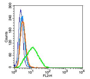 Blank control: RSC96 cells (blue).
Primary Antibody:Rabbit Anti-CD9 antibody(SL2489R), Dilution: 1μg in 100 μL 1X PBS containing 0.5% BSA;
Isotype Control Antibody: Rabbit IgG(orange) ,used under the same conditions );
Secondary Antibody: Goat anti-rabbit IgG-PE(white blue), Dilution: 1:200 in 1 X PBS containing 0.5% BSA.
Protocol
Primary antibody were incubated for 30 min on the ice, followed by 1 X PBS containing 0.5% BSA + 10% goat serum (15 min) to block non-specific protein-protein interactions. Then the Goat Anti-rabbit IgG/PE antibody was added into the blocking buffer mentioned above to react with the primary antibody at 1/200 dilution for 30 min on ice. Acquisition of 20,000 events was performed.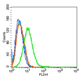 Blank control: Raji(blue).
Primary Antibody: Rabbit Anti-CD9 antibody(SL2489R), Dilution: 5μg in 100 μL 1X PBS containing 0.5% BSA;
Isotype Control Antibody: Rabbit IgG (orange) ,used under the same conditions.
Secondary Antibody: Goat anti-rabbit IgG-PE(white blue), Dilution: 1:200 in 1 X PBS containing 0.5% BSA.
Cartpieces
Totalgoods,subtotals:¥Checkout
References (0)
No References
Bought notes(bought amounts latest0)
No one bought this product
User Comment(Total0User Comment Num)
- No comment


 +86 571 56623320
 +86 18668110335

