
Rabbit Anti-IRAK4 antibody
IL-1 Receptor-associated Kinase 4; 8430405M07Rik; 9330209D03Rik; IPD1; IRAK4; NY-REN-64; REN64; INTERLEUKIN RECEPTOR-ASSOCIATED KINASE 4; Interleukin 1 receptor associated kinase 4 mutant form 1; Interleukin-1 receptor-associated kinase 4; Interleukin1 re
View History [Clear]
Details
Product Name IRAK4 Chinese Name 白介素-1受体相关激酶4抗体 Alias IL-1 Receptor-associated Kinase 4; 8430405M07Rik; 9330209D03Rik; IPD1; IRAK4; NY-REN-64; REN64; INTERLEUKIN RECEPTOR-ASSOCIATED KINASE 4; Interleukin 1 receptor associated kinase 4 mutant form 1; Interleukin-1 receptor-associated kinase 4; Interleukin1 receptor associated kinase 4; IPD1; IRAK 4; IRAK-4; IRAK4 mutated form 1; IRAK4_HUMAN; LOC 51135; NY REN 64 antigen; Renal carcinoma antigen NY-REN-64. literatures Research Area Cell biology immunology Kinases and Phosphatases Immunogen Species Rabbit Clonality Polyclonal React Species Human, Mouse, Rat, (predicted: Dog, Pig, Cow, Horse, Sheep, ) Applications WB=1:500-2000 ELISA=1:5000-10000 IHC-P=1:100-500 IHC-F=1:100-500 IF=1:100-500 (Paraffin sections need antigen repair)
not yet tested in other applications.
optimal dilutions/concentrations should be determined by the end user.Theoretical molecular weight 51kDa Cellular localization cytoplasmic Form Liquid Concentration 1mg/ml immunogen KLH conjugated synthetic peptide derived from human IRAK4: 21-120/460 Lsotype IgG Purification affinity purified by Protein A Buffer Solution 0.01M TBS(pH7.4) with 1% BSA, 0.03% Proclin300 and 50% Glycerol. Storage Shipped at 4℃. Store at -20 °C for one year. Avoid repeated freeze/thaw cycles. Attention This product as supplied is intended for research use only, not for use in human, therapeutic or diagnostic applications. PubMed PubMed Product Detail This gene encodes a kinase that activates NF-kappaB in both the Toll-like receptor (TLR) and T-cell receptor (TCR) signaling pathways. The protein is essential for most innate immune responses. Mutations in this gene result in IRAK4 deficiency and recurrent invasive pneumococcal disease. Multiple transcript variants encoding different isoforms have been found for this gene. [provided by RefSeq, Aug 2011]
Function:
Serine/threonine-protein kinase that plays a critical role in initiating innate immune response against foreign pathogens. Involved in Toll-like receptor (TLR) and IL-1R signaling pathways. Is rapidly recruited by MYD88 to the receptor-signaling complex upon TLR activation to form the Myddosome together with IRAK2. Phosphorylates initially IRAK1, thus stimulating the kinase activity and intensive autophosphorylation of IRAK1. Phosphorylates E3 ubiquitin ligases Pellino proteins (PELI1, PELI2 and PELI3) to promote pellino-mediated polyubiquitination of IRAK1. Then, the ubiquitin-binding domain of IKBKG/NEMO binds to polyubiquitinated IRAK1 bringing together the IRAK1-MAP3K7/TAK1-TRAF6 complex and the NEMO-IKKA-IKKB complex. In turn, MAP3K7/TAK1 activates IKKs (CHUK/IKKA and IKBKB/IKKB) leading to NF-kappa-B nuclear translocation and activation. Alternatively, phosphorylates TIRAP to promote its ubiquitination and subsequent degradation. Phosphorylates NCF1 and regulates NADPH oxidase activation after LPS stimulation suggesting a similar mechanism during microbial infections.
Subunit:
Associates with MYD88 and IRAK2 to form a ternary complex called the Myddosome. Once phosphorylated, IRAK4 dissociates from the receptor complex and then associates with the TNF receptor-associated factor 6 (TRAF6), IRAK1, and PELI1; this intermediate complex is required for subsequent NF-kappa-B activation. Interacts with IL1RL1.
Subcellular Location:
Cytoplasm.
Post-translational modifications:
Phosphorylated.
DISEASE:
Defects in IRAK4 are the cause of recurrent isolated invasive pneumococcal disease type 1 (IPD1) [MIM:610799]. Recurrent invasive pneumococcal disease (IPD) is defined as two episodes of IPD occurring at least 1 month apart, whether caused by the same or different serotypes or strains. Recurrent IPD occurs in at least 2% of patients in most series, making IPD the most important known risk factor for subsequent IPD.
Defects in IRAK4 are the cause of IRAK4 deficiency (IRAK4D) [MIM:607676]. IRAK4 deficiency causes extracellular pyogenic bacterial and fungal infections in otherwise healthy children.
Similarity:
Belongs to the protein kinase superfamily. TKL Ser/Thr protein kinase family. Pelle subfamily.
Contains 1 death domain.
Contains 1 protein kinase domain.
SWISS:
Q9NWZ3
Gene ID:
51135
Database links:Entrez Gene: 51135 Human
Entrez Gene: 266632 Mouse
Omim: 606883 Human
SwissProt: Q9NWZ3 Human
SwissProt: Q8R4K2 Mouse
Unigene: 138499 Human
Unigene: 422858 Mouse
Product Picture 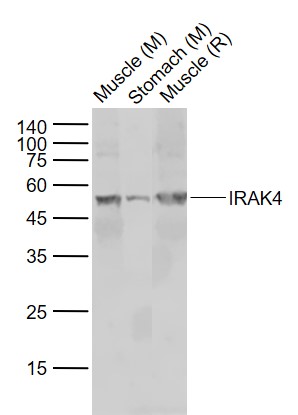 Sample:
Lane 1: Muscle (Mouse) Lysate at 40 ug
Lane 2: Stomach (Mouse) Lysate at 40 ug
Lane 3: Muscle (Rat) Lysate at 40 ug
Primary: Anti-IRAK4 (SL2440R) at 1/1000 dilution
Secondary: IRDye800CW Goat Anti-Rabbit IgG at 1/20000 dilution
Predicted band size: 51-55 kD
Observed band size: 51 kD
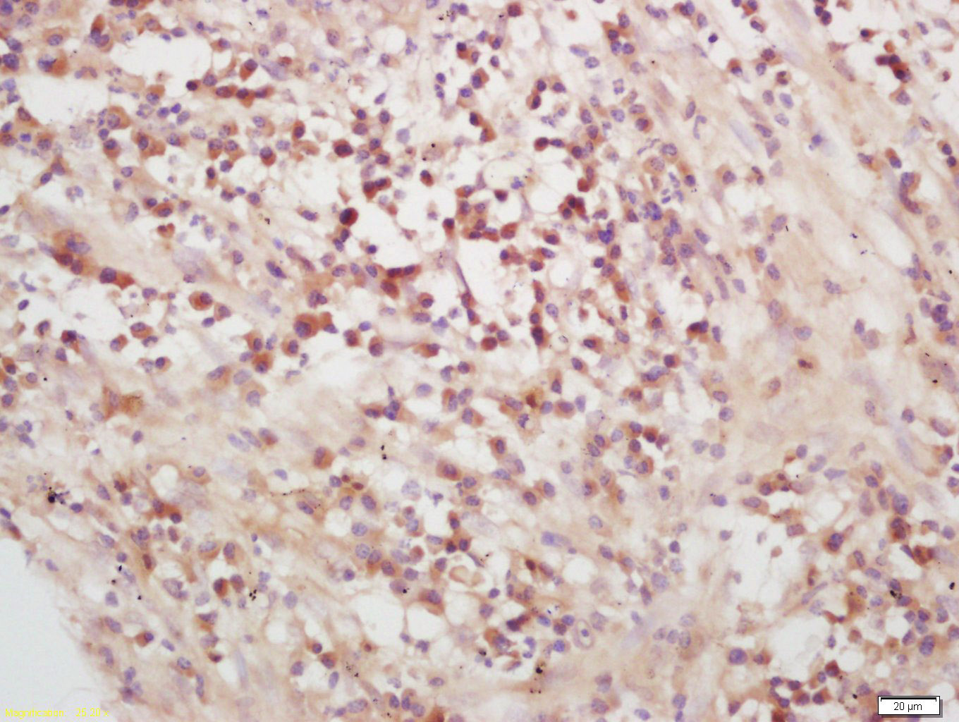 Tissue/cell: human lung carcinoma; 4% Paraformaldehyde-fixed and paraffin-embedded;
Antigen retrieval: citrate buffer ( 0.01M, pH 6.0 ), Boiling bathing for 15min; Block endogenous peroxidase by 3% Hydrogen peroxide for 30min; Blocking buffer (normal goat serum,C-0005) at 37℃ for 20 min;
Incubation: Anti-IRAK4 Polyclonal Antibody, Unconjugated(SL2440R) 1:200, overnight at 4°C, followed by conjugation to the secondary antibody(SP-0023) and DAB(C-0010) staining
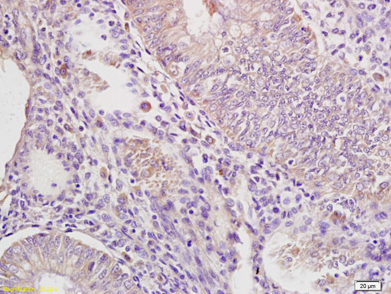 Tissue/cell: human endometrium carcinoma; 4% Paraformaldehyde-fixed and paraffin-embedded;
Antigen retrieval: citrate buffer ( 0.01M, pH 6.0 ), Boiling bathing for 15min; Block endogenous peroxidase by 3% Hydrogen peroxide for 30min; Blocking buffer (normal goat serum,C-0005) at 37℃ for 20 min;
Incubation: Anti-IRAK4 Polyclonal Antibody, Unconjugated(SL2440R) 1:200, overnight at 4°C, followed by conjugation to the secondary antibody(SP-0023) and DAB(C-0010) staining
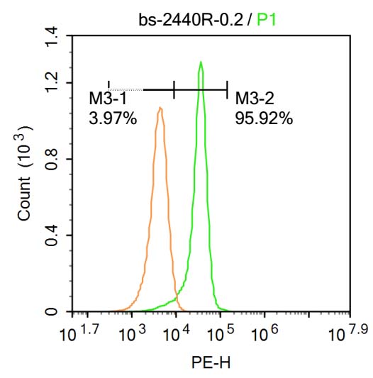 Blank control:Molt-4.
Primary Antibody (green line): Rabbit Anti-IRAK4 antibody (SL2440R)
Dilution: 0.2μg /10^6 cells;
Isotype Control Antibody (orange line): Rabbit IgG .
Secondary Antibody : Goat anti-rabbit IgG-PE
Dilution: 0.2μg /test.
Protocol
The cells were fixed with 4% PFA (10min at room temperature)and then permeabilized with 20% PBST for 20 min at-20℃. The cells were then incubated in 5%BSA to block non-specific protein-protein interactions for 30 min at at room temperature .Cells stained with Primary Antibody for 30 min at room temperature. The secondary antibody used for 40 min at room temperature. Acquisition of 20,000 events was performed.
Cartpieces
Totalgoods,subtotals:¥Checkout
References (0)
No References
Bought notes(bought amounts latest0)
No one bought this product
User Comment(Total0User Comment Num)
- No comment


 +86 571 56623320
 +86 18668110335

