Rabbit Anti-TREM1 antibody
Triggering receptor expressed on myeloid cells 1; TREM 1; TREM-1; Triggering receptor expressed on monocytes 1; Triggering receptor TREM 1; Triggering receptor TREM1; Triggering-receptor TREM 1; Triggering-receptor TREM-1; Triggering-receptor TREM1; TREM1
View History [Clear]
Details
Product Name TREM1 Chinese Name 髓系细胞触发受体1抗体 Alias Triggering receptor expressed on myeloid cells 1; TREM 1; TREM-1; Triggering receptor expressed on monocytes 1; Triggering receptor TREM 1; Triggering receptor TREM1; Triggering-receptor TREM 1; Triggering-receptor TREM-1; Triggering-receptor TREM1; TREM1_HUMAN; CD354. Research Area immunology The cell membrane受体 Immunogen Species Rabbit Clonality Polyclonal React Species Human, Mouse, (predicted: Rat, ) Applications WB=1:500-2000 IHC-P=1:100-500 IHC-F=1:100-500 IF=1:100-500 (Paraffin sections need antigen repair)
not yet tested in other applications.
optimal dilutions/concentrations should be determined by the end user.Theoretical molecular weight 24kDa Cellular localization The cell membrane Secretory protein Form Liquid Concentration 1mg/ml immunogen KLH conjugated synthetic peptide derived from mouse TREM1: 1-100/230 <Extracellular> Lsotype IgG Purification affinity purified by Protein A Buffer Solution 0.01M TBS(pH7.4) with 1% BSA, 0.03% Proclin300 and 50% Glycerol. Storage Shipped at 4℃. Store at -20 °C for one year. Avoid repeated freeze/thaw cycles. Attention This product as supplied is intended for research use only, not for use in human, therapeutic or diagnostic applications. PubMed PubMed Product Detail This gene encodes a receptor belonging to the Ig superfamily that is expressed on myeloid cells. This protein amplifies neutrophil and monocyte-mediated inflammatory responses triggered by bacterial and fungal infections by stimulating release of pro-inflammatory chemokines and cytokines, as well as increased surface expression of cell activation markers. Alternatively spliced transcript variants encoding different isoforms have been noted for this gene.[provided by RefSeq, Jun 2011].
Function:
Stimulates neutrophil and monocyte-mediated inflammatory responses. Triggers release of pro-inflammatory chemokines and cytokines, as well as increased surface expression of cell activation markers. Amplifier of inflammatory responses that are triggered by bacterial and fungal infections and is a crucial mediator of septic shock (By similarity).
Subunit:
Interacts with TYROBP/DAP12.
Subcellular Location:
Isoform 1: Cell membrane; Single-pass type I membrane protein (Potential). Isoform 2: Secreted (Potential).
Tissue Specificity:
Highly expressed in adult liver, lung and spleen than in corresponding fetal tissue. Also expressed in the lymph node, placenta, spinal cord and heart tissues. Expression is more elevated in peripheral blood leukocytes than in the bone marrow and in normal cells than malignant cells. Expressed at low levels in the early development of the hematopoietic system and in the promonocytic stage and at high levels in mature monocytes. Strongly expressed in acute inflammatory lesions caused by bacteria and fungi. Isoform 2 was detected in the lung, liver and mature monocytes.
Similarity:
Contains 1 Ig-like V-type (immunoglobulin-like) domain.
SWISS:
Q9JKE2
Gene ID:
58217
Database links:Entrez Gene: 54210 Human
Entrez Gene: 58217 Mouse
Omim: 605085 Human
SwissProt: Q9NP99 Human
SwissProt: Q9JKE2 Mouse
Unigene: 283022 Human
Unigene: 248352 Mouse
Product Picture
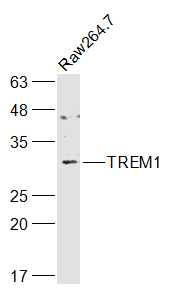
Raw264.7(Mouse) Cell Lysate at 30 ug
Primary: Anti-TREM1 (SL23400R) at 1/1000 dilution
Secondary: IRDye800CW Goat Anti-Rabbit IgG at 1/20000 dilution
Predicted band size: 24 kD
Observed band size: 29 kD
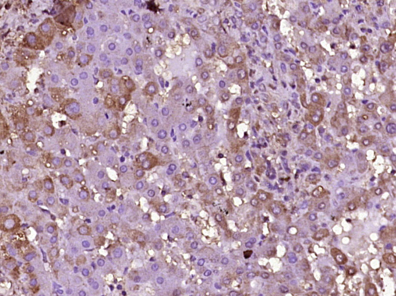
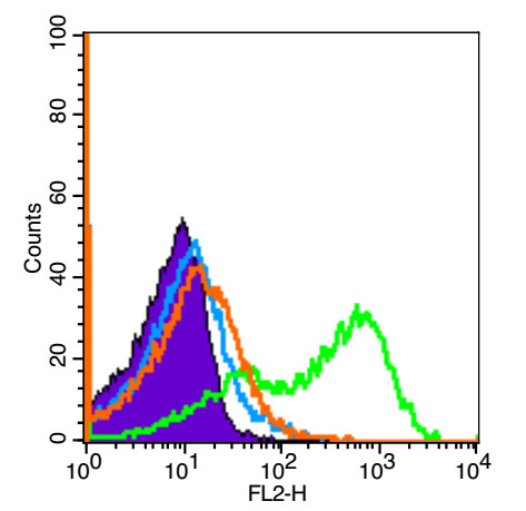
Paraformaldehyde-fixed, paraffin embedded (Human liver); Antigen retrieval by boiling in sodium citrate buffer (pH6.0) for 15min; Block endogenous peroxidase by 3% hydrogen peroxide for 20 minutes; Blocking buffer (normal goat serum) at 37°C for 30min; Antibody incubation with (TREM1) Polyclonal Antibody, Unconjugated (SL23400R) at 1:400 overnight at 4°C, followed by operating according to SP Kit(Rabbit) (sp-0023) instructionsand DAB staining.Blank control (black line): Mouse spleen(Black).
Primary Antibody (green line): Rabbit Anti-TREM1 antibody (SL23400R)
Dilution: 1μg /10^6 cells;
Isotype Control Antibody (orange line): Rabbit IgG .
Secondary Antibody (white blue line): Goat anti-rabbit IgG-PE
Dilution: 3μg /test.
Protocol
The cells were fixed with 4% paraformaldehyde for 10 min at room temperature. The cells were then incubated in 5% BSA to block non-specific protein-protein interactions for 30 min at room temperature. Cells stained with Primary Antibody for 30 min at room temperature The secondary antibody used for 40 min at room temperature. Acquisition of 10,000 events was performed.
Partial purchase records(bought amounts latest0)
No one bought this product
User Comment(Total0User Comment Num)
- No comment


 +86 571 56623320


