
Rabbit Anti-CD33 antibody
CD33_HUMAN; CD33 antigen; CD33 molecule; FLJ00391; gp67; Myeloid cell surface antigen CD33; Myeloid cell surface antigen CD33 precursor; p67; Sialic acid binding Ig like lectin 3; Sialic acid binding immunoglobulin like lectin 3; SIGLEC3; Siglec-3.
View History [Clear]
Details
Product Name CD33 Chinese Name CD33抗体 Alias CD33_HUMAN; CD33 antigen; CD33 molecule; FLJ00391; gp67; Myeloid cell surface antigen CD33; Myeloid cell surface antigen CD33 precursor; p67; Sialic acid binding Ig like lectin 3; Sialic acid binding immunoglobulin like lectin 3; SIGLEC3; Siglec-3. literatures Research Area immunology Neurobiology Stem cells Immunogen Species Rabbit Clonality Polyclonal React Species Human, Applications WB=1:500-2000 ELISA=1:5000-10000 IHC-P=1:100-500 IHC-F=1:100-500 IF=1:100-500 (Paraffin sections need antigen repair)
not yet tested in other applications.
optimal dilutions/concentrations should be determined by the end user.Theoretical molecular weight 40/25kDa Cellular localization The cell membrane Form Liquid Concentration 1mg/ml immunogen KLH conjugated synthetic peptide derived from human CD33: 261-364/364 <Cytoplasmic> Lsotype IgG Purification affinity purified by Protein A Buffer Solution 0.01M TBS(pH7.4) with 1% BSA, 0.03% Proclin300 and 50% Glycerol. Storage Shipped at 4℃. Store at -20 °C for one year. Avoid repeated freeze/thaw cycles. Attention This product as supplied is intended for research use only, not for use in human, therapeutic or diagnostic applications. PubMed PubMed Product Detail Enables protein phosphatase binding activity and sialic acid binding activity. Involved in several processes, including negative regulation of cytokine production; negative regulation of monocyte activation; and positive regulation of protein tyrosine phosphatase activity. Located in several cellular components, including Golgi apparatus; external side of plasma membrane; and peroxisome. [provided by Alliance of Genome Resources, Apr 2022]
Function:
Putative adhesion molecule of myelomonocytic-derived cells that mediates sialic-acid dependent binding to cells. Preferentially binds to alpha-2,6-linked sialic acid. The sialic acid recognition site may be masked by cis interactions with sialic acids on the same cell surface. In the immune response, may act as an inhibitory receptor upon ligand induced tyrosine phosphorylation by recruiting cytoplasmic phosphatase(s) via their SH2 domain(s) that block signal transduction through dephosphorylation of signaling molecules. Induces apoptosis in acute myeloid leukemia (in vitro).
Subunit:
Interacts with PTPN6/SHP-1 and PTPN11/SHP-2 upon phosphorylation.
Subcellular Location:
Cell membrane; Single-pass type I membrane protein.
Tissue Specificity:
Monocytic/myeloid lineage cells.
Post-translational modifications:
Phosphorylation of Tyr-340 is involved in binding to PTPN6 and PTPN11. Phosphorylation of Tyr-358 is involved in binding to PTPN6.
Similarity:
Belongs to the immunoglobulin superfamily. SIGLEC (sialic acid binding Ig-like lectin) family.
Contains 1 Ig-like C2-type (immunoglobulin-like) domain.
Contains 1 Ig-like V-type (immunoglobulin-like) domain.
SWISS:
P20138
Gene ID:
945
Database links:Entrez Gene: 945 Human
Entrez Gene: 12489 Mouse
Omim: 159590 Human
SwissProt: P20138 Human
SwissProt: Q63994 Mouse
Unigene: 83731 Human
Unigene: 140157 Mouse
CD33(Siglec-3)也是急性lymphocyte白血病抗原,细胞表面跨膜glycoprotein.CD33表达于早期Blymphocyte,某些粒性白细胞,骨髓基质细胞,部分上皮组织及其起源的Tumour细胞中也有表达.主要应用于某些恶性淋巴瘤和白血病的分型.Product Picture 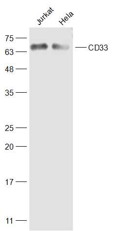 Sample:
Jurkat(Human) Cell Lysate at 30 ug
Hela(Human) Cell Lysate at30 ug
Primary: Anti-CD33 (SL2042R) at 1/300 dilution
Secondary: IRDye800CW Goat Anti-Rabbit IgG at 1/20000 dilution
Predicted band size: 40/25 kD
Observed band size: 67 kD
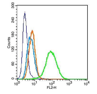 Blank control: U937 (blue).
Primary Antibody:Rabbit Anti- CD33 antibody(SL2042R), Dilution: 1μg in 100 μL 1X PBS containing 0.5% BSA;
Isotype Control Antibody: Rabbit IgG(orange) ,used under the same conditions );
Secondary Antibody: Goat anti-rabbit IgG-PE(white blue), Dilution: 1:200 in 1 X PBS containing 0.5% BSA.
Protocol
The cells were fixed with 2% paraformaldehyde (10 min). Primary antibody (SL2042R, 1μg /1x10^6 cells) were incubated for 30 min on the ice, followed by 1 X PBS containing 0.5% BSA + 1 0% goat serum (15 min) to block non-specific protein-protein interactions. Then the Goat Anti-rabbit IgG/PE antibody was added into the blocking buffer mentioned above to react with the primary antibody at 1/200 dilution for 30 min on ice. Acquisition of 20,000 events was performed.
Cartpieces
Totalgoods,subtotals:¥Checkout
Bought notes(bought amounts latest0)
No one bought this product
User Comment(Total0User Comment Num)
- No comment


 +86 571 56623320
 +86 18668110335

