
Rabbit Anti-LC3C antibody
LC3 like protein 2; MAP1 light chain 3 like protein 3; Microtubule-associated proteins 1A/1B light chain 3C; Autophagy-related protein LC3 C; Autophagy-related ubiquitin-like modifier LC3 C; MAP1 light chain 3-like protein 3; MAP1A/MAP1B light chain 3 C;
View History [Clear]
Details
Product Name LC3C Chinese Name 微管相关蛋白轻链3C抗体 Alias LC3 like protein 2; MAP1 light chain 3 like protein 3; Microtubule-associated proteins 1A/1B light chain 3C; Autophagy-related protein LC3 C; Autophagy-related ubiquitin-like modifier LC3 C; MAP1 light chain 3-like protein 3; MAP1A/MAP1B light chain 3 C; MAP1A/MAP1B LC3 C; Microtubule-associated protein 1 light chain 3 gamma; MAP1LC3C; MLP3C_HUMAN. literatures Research Area Cardiovascular Signal transduction Cytoskeleton Autophagy Immunogen Species Rabbit Clonality Polyclonal React Species Human, (predicted: Mouse, ) Applications ELISA=1:5000-10000 Flow-Cyt=1μg/Test
not yet tested in other applications.
optimal dilutions/concentrations should be determined by the end user.Theoretical molecular weight 16kDa Cellular localization cytoplasmic The cell membrane Form Liquid Concentration 1mg/ml immunogen KLH conjugated synthetic peptide derived from human LC3C: 1-50/147 Lsotype IgG Purification affinity purified by Protein A Buffer Solution Preservative: 15mM Sodium Azide, Constituents: 1% BSA, 0.01M PBS, pH 7.4 Storage Shipped at 4℃. Store at -20 °C for one year. Avoid repeated freeze/thaw cycles. Attention This product as supplied is intended for research use only, not for use in human, therapeutic or diagnostic applications. PubMed PubMed Product Detail A major contributor to cellular homeostasis is the ability of the cell to strike a balance between the formation and degradation/removal of its cellular components. This process of internal cellular turn-over is called autophagy (self-eating), and is facilitated by a pathway of around 16 interacting proteins in the human. LC3, a ubiquitin-like modifier protein, is the human homolog of yeast Apg8 and is involved in the formation of autophagosomal vacuoles, called autophagosomes. There are three isoforms of human LC3 (named MAP1LC3A, MAP1LC3B, and MAP1LC3C), which exhibit different tissue distributions. A disruption to the autophagic process is now associated with the progression of several cancers, neurodegenerative disorders and cardiac pathologies, where LC3 is widely employed as a marker for autophagy.
Function:
Probably involved in formation of autophagosomal vacuoles (autophagosomes).
Subunit:
3 different light chains, LC1, LC2 and LC3, can associate with MAP1A and MAP1B proteins.
Subcellular Location:
Cytoplasm, cytoskeleton. Endomembrane system; Lipid-anchor. Cytoplasmic vesicle, autophagosome membrane; Lipid-anchor.
Tissue Specificity:
Most abundant in placenta, lung and ovary.
Similarity:
Belongs to the MAP1 LC3 family.
SWISS:
Q9BXW4
Gene ID:
440738
Database links:
Entrez Gene: 440738 Human
Omim: 609605 Human
SwissProt: Q9BXW4 Human
Unigene: 534971 Human
Product Picture 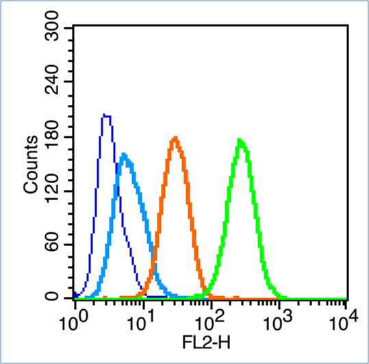 Blank control (blue line): Hela (fixed with 70% ethanol (Overmight at 4℃) and then permeabilized with 90% ice-cold methanol for 30 min at -20℃).
Primary Antibody (green line): Rabbit Anti-LC3C antibody (SL20416R),Dilution: 1μg /10^6 cells;
Isotype Control Antibody (orange line): Rabbit IgG .
Secondary Antibody (white blue line): Goat anti-rabbit IgG-PE,Dilution: 1μg /test.
 Blank control:293T.
Primary Antibody (green line): Rabbit Anti-LC3C antibody (SL20416R)
Dilution: 1μg /10^6 cells;
Isotype Control Antibody (orange line): Rabbit IgG .
Secondary Antibody : Goat anti-rabbit IgG-AF488
Dilution: 1μg /test.
Protocol
The cells were fixed with 4% PFA (10min at room temperature)and then permeabilized with 0.1% PBST for 20 min at room temperature. The cells were then incubated in 5%BSA to block non-specific protein-protein interactions for 30 min at room temperature .Cells stained with Primary Antibody for 30 min at room temperature. The secondary antibody used for 40 min at room temperature. Acquisition of 20,000 events was performed.
Cartpieces
Totalgoods,subtotals:¥Checkout
Bought notes(bought amounts latest0)
No one bought this product
User Comment(Total0User Comment Num)
- No comment


 +86 571 56623320
 +86 18668110335

