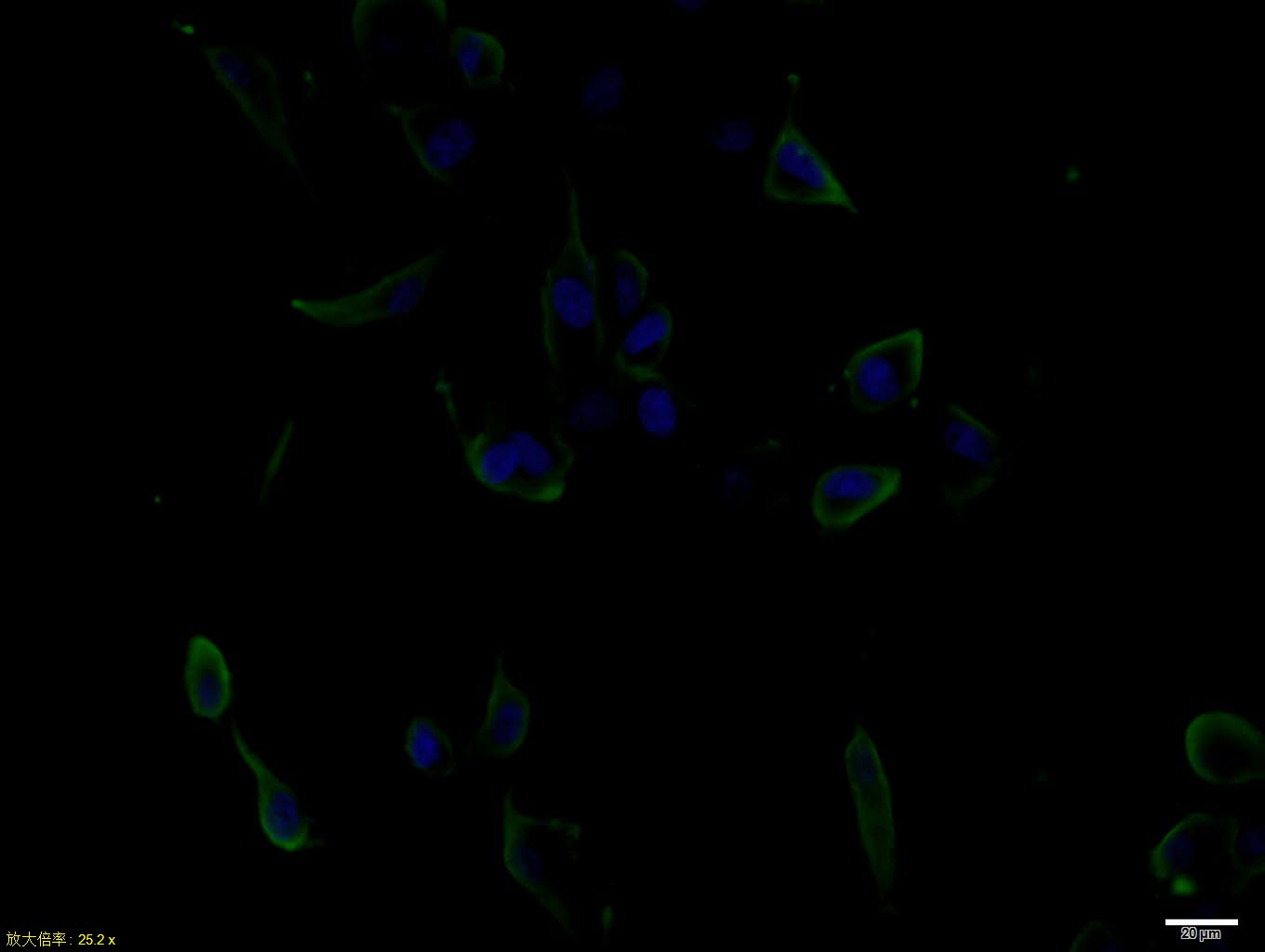
Rabbit Anti-EGF antibody
Beta urogastrone; Epidermal Growth Factor; Pro epidermal growth factor; URG; Urogastrone; EGF; EGF_HUMAN; Epidermal Growth Factor; HOMG4; OTTHUMP00000219721; OTTHUMP00000219722; Pro epidermal growth factor; URG; Urogastrone.
View History [Clear]
Details
Product Name EGF Chinese Name 表皮生长因子抗体 Alias Beta urogastrone; Epidermal Growth Factor; Pro epidermal growth factor; URG; Urogastrone; EGF; EGF_HUMAN; Epidermal Growth Factor; HOMG4; OTTHUMP00000219721; OTTHUMP00000219722; Pro epidermal growth factor; URG; Urogastrone. literatures Research Area Tumour Cell biology immunology Growth factors and hormones Immunogen Species Rabbit Clonality Polyclonal React Species Human, Applications ELISA=1:5000-10000 IHC-P=1:100-500 IHC-F=1:100-500 Flow-Cyt=1ug/Test ICC=1:100 IF=1:100-500 (Paraffin sections need antigen repair)
not yet tested in other applications.
optimal dilutions/concentrations should be determined by the end user.Theoretical molecular weight 5.8/133kDa Cellular localization The cell membrane Form Liquid Concentration 1mg/ml immunogen KLH conjugated synthetic peptide derived from human EGF: 31-53/53 (1007-1023/1207) Lsotype IgG Purification affinity purified by Protein A Buffer Solution 0.01M TBS(pH7.4) with 1% BSA, 0.03% Proclin300 and 50% Glycerol. Storage Shipped at 4℃. Store at -20 °C for one year. Avoid repeated freeze/thaw cycles. Attention This product as supplied is intended for research use only, not for use in human, therapeutic or diagnostic applications. PubMed PubMed Product Detail This gene encodes a member of the epidermal growth factor superfamily. The encoded preproprotein is proteolytically processed to generate the 53-amino acid epidermal growth factor peptide. This protein acts a potent mitogenic factor that plays an important role in the growth, proliferation and differentiation of numerous cell types. This protein acts by binding with high affinity to the cell surface receptor, epidermal growth factor receptor. Defects in this gene are the cause of hypomagnesemia type 4. Dysregulation of this gene has been associated with the growth and progression of certain cancers. Alternative splicing results in multiple transcript variants, at least one of which encodes a preproprotein that is proteolytically processed. [provided by RefSeq, Jan 2016].
Function:
EGF stimulates the growth of various epidermal and epithelial tissues in vivo and in vitro and of some fibroblasts in cell culture. Magnesiotropic hormone that stimulates magnesium reabsorption in the renal distal convoluted tubule via engagement of EGFR and activation of the magnesium channel TRPM6.
Subunit:
Interacts with EGFR and promotes EGFR dimerization. Interacts with RHBDF2. Interacts with RHBDF1; may retain EGF in the endoplasmic reticulum and regulates its degradation through the endoplasmic reticulum-associated degradation (ERAD).
Subcellular Location:
Membrane; Single-pass type I membrane protein.
Tissue Specificity:
Expressed in kidney, salivary gland, cerebrum and prostate.
Post-translational modifications:
Phosphorylation at Ser-695 is partial and occurs only if Thr-693 is phosphorylated. Phosphorylation at Thr-678 and Thr-693 by PRKD1 inhibits EGF-induced MAPK8/JNK1 activation. Dephosphorylation by PTPRJ prevents endocytosis and stabilizes the receptor at the plasma membrane. Autophosphorylation at Tyr-1197 is stimulated by methylation at Arg-1199 and enhances interaction with PTPN6. Autophosphorylation at Tyr-1092 and/or Tyr-1110 recruits STAT3.
Monoubiquitinated and polyubiquitinated upon EGF stimulation; which does not affect tyrosine kinase activity or signaling capacity but may play a role in lysosomal targeting. Polyubiquitin linkage is mainly through 'Lys-63', but linkage through 'Lys-48', 'Lys-11' and 'Lys-29' also occur. Deubiquitinated by OTUD7B, preventing degradation.
Methylated. Methylation at Arg-1199 by PRMT5 positively stimulates phosphorylation at Tyr-1197.
DISEASE:
Defects in EGF are the cause of hypomagnesemia type 4 (HOMG4) [MIM:611718]; also known as renal hypomagnesemia normocalciuric. HOMG4 is a disorder characterized by massive renal hypomagnesemia and normal levels of serum calcium and calcium excretion. Clinical features include seizures, mild-to mederate psychomotor retardation, and brisk tendon reflexes.
Similarity:
Contains 9 EGF-like domains.
Contains 9 LDL-receptor class B repeats.
SWISS:
P01133
Gene ID:
1950
Database links:Entrez Gene: 13645 Mouse
Entrez Gene: 1950 Human
Omim: 131530 Human
SwissProt: P01133 Human
SwissProt: P01132 Mouse
Unigene: 419815 Human
表皮生长因子是一种小肽,由53个氨基酸残基组成, 是类EGF大家族的一个成员,是一种多功能的生长因子,在体内体外都对多种组织细胞有强烈的促分裂作用。EGF同应答细胞表面的特异受体结合,一旦结合,便促进受体二聚化并使细胞质位点磷酸化。被激活的受体至少可与5种具有不同信号序列的蛋白结合,进行Signal transduction,在翻译水平上对蛋白质的合成起调节作用。此外EGF可提高细胞内DNA拓扑异构酶活性,也可促进一些与增殖有关的基因表达,如myc 、fos等。Product Picture
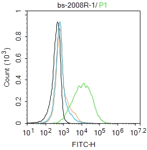
Primary Antibody (green line): Rabbit Anti-EGF antibody (SL2008R)
Dilution: 1μg /10^6 cells;
Isotype Control Antibody (orange line): Rabbit IgG .
Secondary Antibody : Goat anti-rabbit IgG-AF488
Dilution: 1μg /test.
Protocol
The cells were incubated in 5%BSA to block non-specific protein-protein interactions for 30 min at room temperature .Cells stained with Primary Antibody for 30 min at room temperature. The secondary antibody used for 40 min at room temperature. Acquisition of 20,000 events was performed.
Partial purchase records(bought amounts latest0)
No one bought this product
User Comment(Total0User Comment Num)
- No comment


 +86 571 56623320


