
Rabbit Anti-Dopamine D5 receptor antibody
D(1B) dopamine receptor; D(5) dopamine receptor; D1 beta dopamine receptor; D1B dopamine receptor; D1beta dopamine receptor; D5 dopamine receptor; DBDR; Dopamine receptor D1B; DRD 1B; DRD 5; DRD1B; DRD1L2; DRD5; MGC10601; DRD5_HUMAN; DRD5_RAT.
View History [Clear]
Details
Product Name Dopamine D5 receptor Chinese Name 多巴胺受体D5抗体 Alias D(1B) dopamine receptor; D(5) dopamine receptor; D1 beta dopamine receptor; D1B dopamine receptor; D1beta dopamine receptor; D5 dopamine receptor; DBDR; Dopamine receptor D1B; DRD 1B; DRD 5; DRD1B; DRD1L2; DRD5; MGC10601; DRD5_HUMAN; DRD5_RAT. literatures Research Area Tumour Cell biology Neurobiology Signal transduction The cell membrane受体 G protein-coupled receptor G protein signal Immunogen Species Rabbit Clonality Polyclonal React Species Mouse, Rat, (predicted: Human, ) Applications WB=1:500-2000 ELISA=1:5000-10000 IHC-P=1:100-500 IHC-F=1:100-500 Flow-Cyt=1μg /test IF=1:100-500 (Paraffin sections need antigen repair)
not yet tested in other applications.
optimal dilutions/concentrations should be determined by the end user.Theoretical molecular weight 53kDa Cellular localization The cell membrane Form Liquid Concentration 1mg/ml immunogen KLH conjugated synthetic peptide derived from rat DRD5: 251-350/475 Lsotype IgG Purification affinity purified by Protein A Buffer Solution 0.01M TBS(pH7.4) with 1% BSA, 0.03% Proclin300 and 50% Glycerol. Storage Shipped at 4℃. Store at -20 °C for one year. Avoid repeated freeze/thaw cycles. Attention This product as supplied is intended for research use only, not for use in human, therapeutic or diagnostic applications. PubMed PubMed Product Detail This is one of the five types (D1 to D5) of receptors for dopamine. The activity of this receptor is mediated by G proteins which activate adenylyl cyclase. This receptor is neuron specific, localized primarily within limbic regions of the brain. Defects in DRD5 are a cause of blepharospasm and may be a cause of schizophrenia.
Subcellular Location:
Cell membrane; Multi-pass membrane protein.
Tissue Specificity:
Neuron-specific, localized primarily within limbic regions of the brain.
Similarity:
Belongs to the G-protein coupled receptor 1 family.
SWISS:
P25115
Gene ID:
25195
Database links:Entrez Gene: 1816 Human
Entrez Gene: 13492 Mouse
Omim: 126453 Human
SwissProt: P21918 Human
SwissProt: Q8BLD9 Mouse
Unigene: 380681 Human
Unigene: 167154 Mouse
Unigene: 138110 Rat
Product Picture 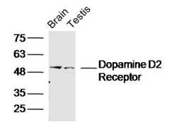 Sample:
Brain (Mouse) Lysate at 40 ug
Testis (Mouse) Lysate at 40 ug
Primary: Anti- Dopamine D5 receptor (SL1747R)at 1/300 dilution
Secondary: IRDye800CW Goat Anti-Rabbit IgG at 1/20000 dilution
Predicted band size: 53kD
Observed band size: 53kD
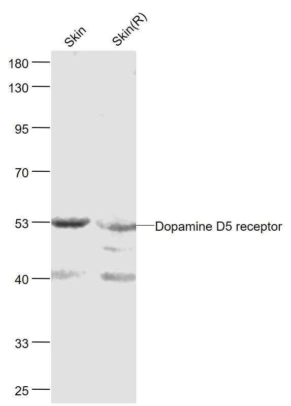 Sample:
Skin(Mouse) Lysate at 40 ug
Skin(Rat) Lysate at 40 ug
Primary: Anti-Dopamine D5 receptor (SL1747R) at 1/300 dilution
Secondary: IRDye800CW Goat Anti-Rabbit IgG at 1/20000 dilution
Predicted band size: 53 kD
Observed band size: 53 kD
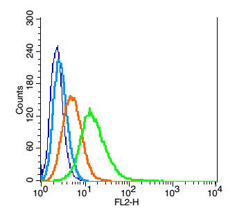 Blank control: RSC96(blue).
Primary Antibody:Rabbit Anti- DRD5 antibody(SL1747R), Dilution: 1μg in 100 μL 1X PBS containing 0.5% BSA;
Isotype Control Antibody: Rabbit IgG(orange) ,used under the same conditions );
Secondary Antibody: Goat anti-rabbit IgG-PE(white blue), Dilution: 1:200 in 1 X PBS containing 0.5% BSA.
Protocol
The cells were fixed with 2% paraformaldehyde (10 min) . Antibody (SL1747R, 1μg /1x10^6 cells) were incubated for 30 min on the ice, followed by 1 X PBS containing 0.5% BSA + 1 0% goat serum (15 min) to block non-specific protein-protein interactions. Then the Goat Anti-rabbit IgG/PE antibody was added into the blocking buffer mentioned above to react with the primary antibody of SL1747R at 1/200 dilution for 30 min on ice. Acquisition of 20,000 events was performed.
Cartpieces
Totalgoods,subtotals:¥Checkout
Bought notes(bought amounts latest0)
No one bought this product
User Comment(Total0User Comment Num)
- No comment


 +86 571 56623320
 +86 18668110335

