
Rabbit Anti-phospho-GAP43 (Ser41)antibody
GAP43 (phospho S41); p-GAP43 (phospho S41); Phospho-GAP43(pSer41); GAP43 (Phospho-Ser41); p-GAP43 (Ser41); p-GAP43 (S41); Growth Associated Protein-43; Neuromodulin; Axonal membrane protein GAP 43; B-50; F1; GAP 43; Growth Associated Protein 43; Nerve Gro
View History [Clear]
Details
Product Name phospho-GAP43 (Ser41) Chinese Name 磷酸化神经生长相关蛋白43抗体 Alias GAP43 (phospho S41); p-GAP43 (phospho S41); Phospho-GAP43(pSer41); GAP43 (Phospho-Ser41); p-GAP43 (Ser41); p-GAP43 (S41); Growth Associated Protein-43; Neuromodulin; Axonal membrane protein GAP 43; B-50; F1; GAP 43; Growth Associated Protein 43; Nerve Growth Related Peptide; Neural phosphoprotein B 50; Neuromodulin; GAP-43; pp46; NEUM_HUMAN; Protein F1; QtrA-11580; QtrA-13071. Product Type Phosphorylated anti Research Area Cell biology immunology Neurobiology Signal transduction Apoptosis transcriptional regulatory factor Immunogen Species Rabbit Clonality Polyclonal React Species Human, Mouse, Rat, (predicted: Dog, ) Applications ELISA=1:5000-10000 IHC-P=1:100-500 IHC-F=1:100-500 Flow-Cyt=0.2g /test IF=1:100-500 (Paraffin sections need antigen repair)
not yet tested in other applications.
optimal dilutions/concentrations should be determined by the end user.Theoretical molecular weight 46kDa Cellular localization cytoplasmic The cell membrane Extracellular matrix Form Liquid Concentration 1mg/ml immunogen KLH conjugated Synthesised phosphopeptide derived from human GAP43 around the phosphorylation site of Ser41: QA(p-S)FR Lsotype IgG Purification affinity purified by Protein A Buffer Solution 0.01M TBS(pH7.4) with 1% BSA, 0.03% Proclin300 and 50% Glycerol. Storage Shipped at 4℃. Store at -20 °C for one year. Avoid repeated freeze/thaw cycles. Attention This product as supplied is intended for research use only, not for use in human, therapeutic or diagnostic applications. PubMed PubMed Product Detail The protein encoded by this gene has been termed a 'growth' or 'plasticity' protein because it is expressed at high levels in neuronal growth cones during development and axonal regeneration. This protein is considered a crucial component of an effective regenerative response in the nervous system. Alternatively spliced transcript variants encoding distinct isoforms have been found for this gene. [provided by RefSeq, Jul 2008]
Function:
This protein is associated with nerve growth. It is a major component of the motile 'growth cones' that form the tips of elongating axons. Plays a role in axonal and dendritic filopodia induction.
Subunit:
Identified in a complex containing FGFR4, NCAM1, CDH2, PLCG1, FRS2, SRC, SHC1, GAP43 and CTTN. Binds calmodulin with a greater affinity in the absence of Ca(2+) than in its presence.
Subcellular Location:
Cell membrane; Peripheral membrane protein; Cytoplasmic side. Cell projection, growth cone membrane; Peripheral membrane protein; Cytoplasmic side. Cell junction, synapse. Cell projection, filopodium membrane; Peripheral membrane protein. Note=Cytoplasmic surface of growth cone and synaptic plasma membranes.
Post-translational modifications:
Phosphorylated at Ser-41 by PHK. Phosphorylation of this protein by a protein kinase C is specifically correlated with certain forms of synaptic plasticity.
Palmitoylation by ARF6 is essential for plasma membrane association and axonal and dendritic filopodia induction. Deacylated by LYPLA2.
Similarity:
Belongs to the neuromodulin family.
Contains 1 IQ domain.
SWISS:
P17677
Gene ID:
2596
Database links:Entrez Gene: 2596 Human
Entrez Gene: 14432 Mouse
GenBank: NP_002036 Human
Omim: 162060 Human
SwissProt: P17677 Human
SwissProt: P06837 Mouse
Unigene: 134974 Human
Unigene: 1222 Mouse
Unigene: 10928 Rat
Product Picture 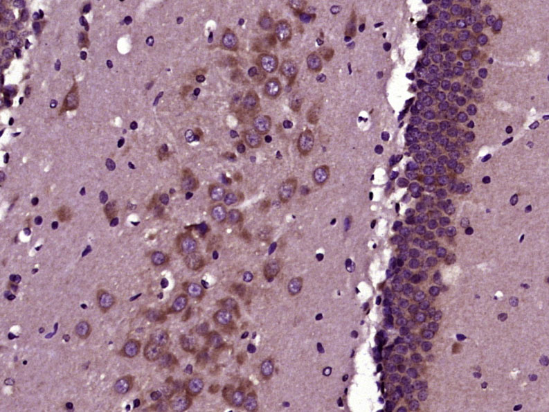 Paraformaldehyde-fixed, paraffin embedded (Rat brain); Antigen retrieval by boiling in sodium citrate buffer (pH6.0) for 15min; Block endogenous peroxidase by 3% hydrogen peroxide for 20 minutes; Blocking buffer (normal goat serum) at 37°C for 30min; Antibody incubation with (phospho-GAP43 (Ser41)) Polyclonal Antibody, Unconjugated (SL1641R) at 1:400 overnight at 4°C, followed by operating according to SP Kit(Rabbit) (sp-0023) instructionsand DAB staining.
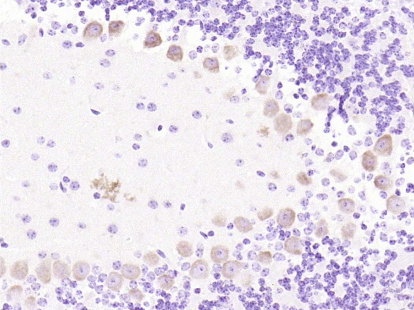 Paraformaldehyde-fixed, paraffin embedded (mouse cerebellum); Antigen retrieval by boiling in sodium citrate buffer (pH6.0) for 15min; Block endogenous peroxidase by 3% hydrogen peroxide for 20 minutes; Blocking buffer (normal goat serum) at 37°C for 30min; Antibody incubation with (phospho-GAP43 (Ser41)) Polyclonal Antibody, Unconjugated (SL1641R) at 1:200 overnight at 4°C, followed by operating according to SP Kit(Rabbit) (sp-0023) instructionsand DAB staining.
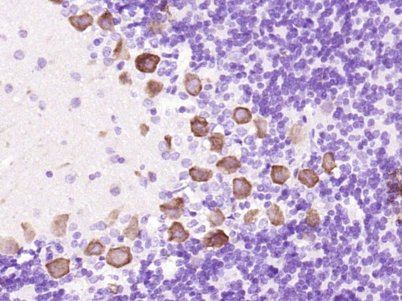 Paraformaldehyde-fixed, paraffin embedded (rat cerebellum); Antigen retrieval by boiling in sodium citrate buffer (pH6.0) for 15min; Block endogenous peroxidase by 3% hydrogen peroxide for 20 minutes; Blocking buffer (normal goat serum) at 37°C for 30min; Antibody incubation with (phospho-GAP43 (Ser41)) Polyclonal Antibody, Unconjugated (SL1641R) at 1:200 overnight at 4°C, followed by operating according to SP Kit(Rabbit) (sp-0023) instructionsand DAB staining.
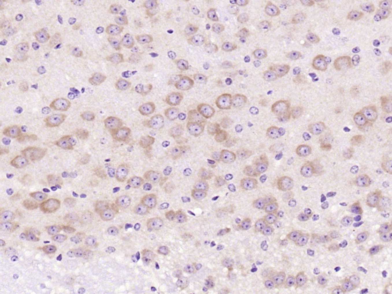 Paraformaldehyde-fixed, paraffin embedded (mouse brain); Antigen retrieval by boiling in sodium citrate buffer (pH6.0) for 15min; Block endogenous peroxidase by 3% hydrogen peroxide for 20 minutes; Blocking buffer (normal goat serum) at 37°C for 30min; Antibody incubation with (phospho-GAP43 (Ser41)) Polyclonal Antibody, Unconjugated (SL1641R) at 1:200 overnight at 4°C, followed by operating according to SP Kit(Rabbit) (sp-0023) instructionsand DAB staining.
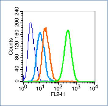 Blank control (blue line): Hela cells (blue).
Primary Antibody (green line): Rabbit Anti-phospho-GAP43 (Ser41) antibody (SL1641R)
Dilution: 0.2μg /10^6 cells;
Isotype Control Antibody (orange line): Rabbit IgG .
Secondary Antibody (white blue line): Goat anti-rabbit IgG-PE
Dilution: 1μg /test.
Protocol
The cells were fixed with 70% methanol (Overnight at 4℃) and then permeabilized with 90% ice-cold methanol for 20 min at -20℃. Cells stained with Primary Antibody for 30 min at room temperature. The cells were then incubated in 1 X PBS/2%BSA/10% goat serum to block non-specific protein-protein interactions followed by the antibody for 15 min at room temperature. The secondary antibody used for 40 min at room temperature. Acquisition of 20,000 events was performed.
Cartpieces
Totalgoods,subtotals:¥Checkout
Bought notes(bought amounts latest0)
No one bought this product
User Comment(Total0User Comment Num)
- No comment


 +86 571 56623320
 +86 18668110335

