
Rabbit Anti-Annexin A1 antibody
Annexin I; AnnexinA1; AnnexinI; ANX 1; ANX A1; ANX1; ANXA 1; ANXA1; ANXA1 protein; Calpactin II; CalpactinII; Chromobindin 9; Chromobindin9; HGNC:533; Lipocortin 1; Lipocortin I; Lipocortin1; LipocortinI; LPC 1; LPC1; p35; Phospholipase A2 inhibitory prot
View History [Clear]
Details
Product Name Annexin A1 Chinese Name 膜粘连蛋白A1抗体 Alias Annexin I; AnnexinA1; AnnexinI; ANX 1; ANX A1; ANX1; ANXA 1; ANXA1; ANXA1 protein; Calpactin II; CalpactinII; Chromobindin 9; Chromobindin9; HGNC:533; Lipocortin 1; Lipocortin I; Lipocortin1; LipocortinI; LPC 1; LPC1; p35; Phospholipase A2 inhibitory protein; ANXA1_HUMAN. literatures Research Area Tumour Immunogen Species Rabbit Clonality Polyclonal React Species Human, Rat, (predicted: Mouse, Pig, Cow, Horse, Rabbit, ) Applications WB=1:500-2000 ELISA=1:5000-10000 IHC-P=1:100-500 IHC-F=1:100-500 ICC=1:100 IF=1:100-500 (Paraffin sections need antigen repair)
not yet tested in other applications.
optimal dilutions/concentrations should be determined by the end user.Theoretical molecular weight 39kDa Cellular localization The nucleus cytoplasmic The cell membrane Form Liquid Concentration 1mg/ml immunogen KLH conjugated synthetic peptide derived from human Annexin A1: 281-346/346 Lsotype IgG Purification affinity purified by Protein A Buffer Solution 0.01M TBS(pH7.4) with 1% BSA, 0.03% Proclin300 and 50% Glycerol. Storage Shipped at 4℃. Store at -20 °C for one year. Avoid repeated freeze/thaw cycles. Attention This product as supplied is intended for research use only, not for use in human, therapeutic or diagnostic applications. PubMed PubMed Product Detail This gene encodes a membrane-localized protein that binds phospholipids. This protein inhibits phospholipase A2 and has anti-inflammatory activity. Loss of function or expression of this gene has been detected in multiple tumors. [provided by RefSeq, Dec 2014]
Function:
Calcium/phospholipid-binding protein which promotes membrane fusion and is involved in exocytosis. This protein regulates phospholipase A2 activity. It seems to bind from two to four calcium ions with high affinity.
Subunit:
Homodimer in placenta (20%); linked by transglutamylation. Interacts with DYSF.
Subcellular Location:
Nucleus. Cytoplasm. Cell projection, cilium. Basolateral cell membrane. Note=Found in the cilium, nucleus and basolateral cell membrane of ciliated cells in the tracheal endothelium. Found in the cytoplasm of type II pneumocytes and alveolar macrophages.
Post-translational modifications:
Phosphorylated by protein kinase C, epidermal growth factor receptor/kinase and TRPM7. Phosphorylation results in loss of the inhibitory activity.
Similarity:
Belongs to the annexin family.
Contains 4 annexin repeats.
SWISS:
P04083
Gene ID:
301
Database links:Entrez Gene: 301 HumanEntrez Gene: 16952 Mouse
SwissProt: P04083 Human
SwissProt: P10107 Mouse
Product Picture 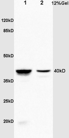 Sample:
Lane1: Kidney(Rat) Lysate at 30 ug
Lane2: Brain(Rat) Lysate at 30 ug
Primary: Anti-Annexin A1 (SL1562R) at 1:200 dilution;
Secondary: HRP conjugated Goat Anti-Rabbit IgG(SL0295G-HRP) at 1: 3000 dilution;
Predicted band size : 39kD
Observed band size : 40kD
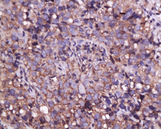 Tissue/cell: human cervical carcinoma; 4% Paraformaldehyde-fixed and paraffin-embedded;
Antigen retrieval: citrate buffer ( 0.01M, pH 6.0 ), Boiling bathing for 15min; Block endogenous peroxidase by 3% Hydrogen peroxide for 30min; Blocking buffer (normal goat serum,C-0005) at 37鈩� for 20 min;
Incubation: Anti-Annexin A1 Polyclonal Antibody, Unconjugated(SL1562R) 1:200, overnight at 4掳C, followed by conjugation to the secondary antibody(SP-0023) and DAB(C-0010) staining
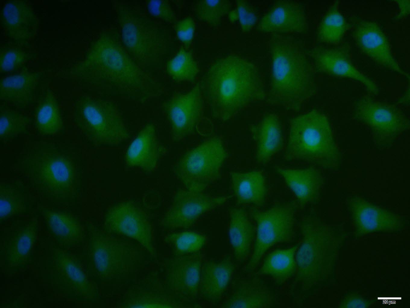 A549 cell; 4% Paraformaldehyde-fixed; Triton X-100 at room temperature for 20 min; Blocking buffer (normal goat serum, C-0005) at 37°C for 20 min; Antibody incubation with (Annexin A1) polyclonal Antibody, Unconjugated (SL1562R) 1:100, 90 minutes at 37°C; followed by a conjugated Goat Anti-Rabbit IgG antibody at 37°C for 90 minutes, DAPI (blue, C02-04002) was used to stain the cell nuclei.
Cartpieces
Totalgoods,subtotals:¥Checkout
Bought notes(bought amounts latest0)
No one bought this product
User Comment(Total0User Comment Num)
- No comment


 +86 571 56623320
 +86 18668110335

