
Rabbit Anti-TEM8 antibody
Anthrax toxin receptor 1; Anthrax toxin receptor 1 precursor; ANTXR 1; ANTXR1; ATR; TEM-8; TEM 8; Tumor Endothelial Marker 8; Tumor endothelial marker 8 precursor; ANTR1_HUMAN; Anthrax toxin receptor-1; FLJ11298; FLJ21776.
View History [Clear]
Details
Product Name TEM8 Chinese Name Tumour血管内皮Maker8抗体 Alias Anthrax toxin receptor 1; Anthrax toxin receptor 1 precursor; ANTXR 1; ANTXR1; ATR; TEM-8; TEM 8; Tumor Endothelial Marker 8; Tumor endothelial marker 8 precursor; ANTR1_HUMAN; Anthrax toxin receptor-1; FLJ11298; FLJ21776. literatures Research Area Cardiovascular Cell biology Signal transduction vascular endothelial cell Immunogen Species Rabbit Clonality Polyclonal React Species Human, Mouse, (predicted: Rat, Rabbit, ) Applications WB=1:500-2000 ELISA=1:5000-10000 IHC-P=1:100-500 IHC-F=1:100-500 ICC=1:100-500 IF=1:100-500 (Paraffin sections need antigen repair)
not yet tested in other applications.
optimal dilutions/concentrations should be determined by the end user.Theoretical molecular weight 63kDa Cellular localization The cell membrane Form Liquid Concentration 1mg/ml immunogen KLH conjugated synthetic peptide derived from human TEM8: 201-300/564 <Extracellular> Lsotype IgG Purification affinity purified by Protein A Buffer Solution 0.01M TBS(pH7.4) with 1% BSA, 0.03% Proclin300 and 50% Glycerol. Storage Shipped at 4℃. Store at -20 °C for one year. Avoid repeated freeze/thaw cycles. Attention This product as supplied is intended for research use only, not for use in human, therapeutic or diagnostic applications. PubMed PubMed Product Detail This gene encodes a type I transmembrane protein and is a tumor-specific endothelial marker that has been implicated in colorectal cancer. The encoded protein has been shown to also be a docking protein or receptor for Bacillus anthracis toxin, the causative agent of the disease, anthrax. The binding of the protective antigen (PA) component, of the tripartite anthrax toxin, to this receptor protein mediates delivery of toxin components to the cytosol of cells. Once inside the cell, the other two components of anthrax toxin, edema factor (EF) and lethal factor (LF) disrupt normal cellular processes. Three alternatively spliced variants that encode different protein isoforms have been described. [provided by RefSeq, Oct 2008]
Function:
Plays a role in cell attachment and migration. Interacts with extracellular matrix proteins and with the actin cytoskeleton. Mediates adhesion of cells to type 1 collagen and gelatin, reorganization of the actin cytoskeleton and promotes cell spreading. Plays a role in the angiogenic response of cultured umbilical vein endothelial cells.
Subunit:
Interacts with gelatin and type 1 collagen. Interacts with the actin cytoskeleton. Binds to the protective antigen (PA) of Bacillus anthracis. Binding does not occur in the presence of calcium.
Subcellular Location:
Cell membrane; Single-pass type I membrane protein. Cell projection, lamellipodium membrane; Single-pass type I membrane protein. Cell projection, filopodium membrane; Single-pass type I membrane protein. Note=At the membrane of lamellipodia and at the tip of actin-enriched filopodia. Colocalizes with actin at the base of lamellipodia.
Tissue Specificity:
Detected in umbilical vein endothelial cells (at protein level). Highly expressed in tumor endothelial cells.
DISEASE:
Defects in ANTXR1 are associated with susceptibility to hemangioma capillary infantile (HCI) [MIM:602089]. HCI are benign, highly proliferative lesions involving aberrant localized growth of capillary endothelium. They are the most common tumor of infancy, occurring in up to 10% of all births. Hemangiomas tend to appear shortly after birth and show rapid neonatal growth for up to 12 months characterized by endothelial hypercellularity and increased numbers of mast cells. This phase is followed by slow involution at a rate of about 10% per year and replacement by fibrofatty stroma
Similarity:
Belongs to the ATR family.
Contains 1 VWFA domain.
SWISS:
Q9H6X2
Gene ID:
84168
Database links:Entrez Gene: 84168 Human
Entrez Gene: 69538 Mouse
Omim: 606410 Human
SwissProt: Q9H6X2 Human
SwissProt: Q9CZ52 Mouse
Unigene: 165859 Human
Unigene: 232525 Mouse
Unigene: 41192 Rat
Product Picture 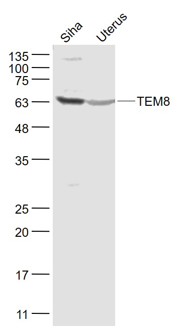 Sample:
Siha(Human) Cell Lysate at 30 ug
Uterus (Mouse) Lysate at 40 ug
Primary: Anti- TEM8 (SL15583R) at 1/1000 dilution
Secondary: IRDye800CW Goat Anti-Rabbit IgG at 1/20000 dilution
Predicted band size: 59 kD
Observed band size: 62 kD
 Sample:
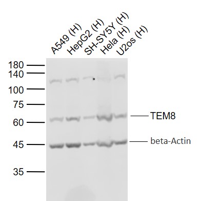
Lane 1: Human A549 cell lysates
Lane 2: Human HepG2 cell lysates
Lane 3: Human SH-SY5Y cell lysates
Lane 4: Human Hela cell lysates
Lane 5: Human U2os cell lysates
Primary: Anti-TEM8 (SL15583R) at 1/1000 dilution
Secondary: IRDye800CW Goat Anti-Rabbit IgG at 1/20000 dilution
Predicted band size: 63 kD
Observed band size: 61 kD
 Sample:
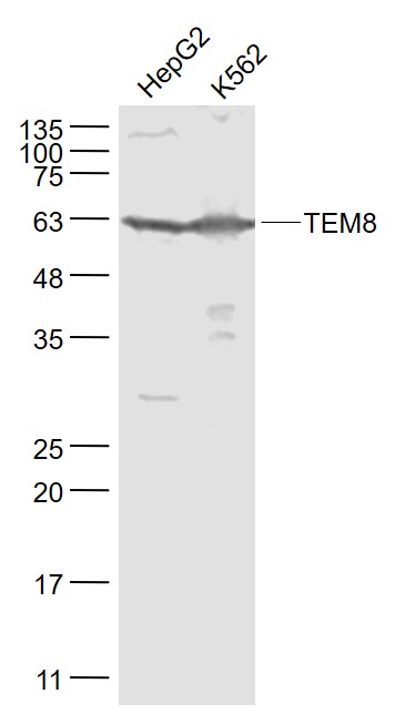
HepG2(Human) Cell Lysate at 30 ug
K562(Human) Cell Lysate at 30 ug
Primary: Anti- TEM8 (SL15583R) at 1/1000 dilution
Secondary: IRDye800CW Goat Anti-Rabbit IgG at 1/20000 dilution
Predicted band size: 59 kD
Observed band size: 62 kD
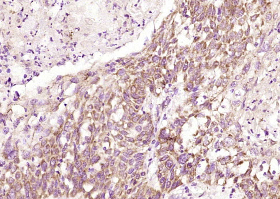 Paraformaldehyde-fixed, paraffin embedded (Human esophageal cancer); Antigen retrieval by boiling in sodium citrate buffer (pH6.0) for 15min; Block endogenous peroxidase by 3% hydrogen peroxide for 20 minutes; Blocking buffer (normal goat serum) at 37°C for 30min; Antibody incubation with (TEM8) Polyclonal Antibody, Unconjugated (SL15583R) at 1:200 overnight at 4°C, followed by operating according to SP Kit(Rabbit) (sp-0023) instructionsand DAB staining.
Cartpieces
Totalgoods,subtotals:¥Checkout
Bought notes(bought amounts latest0)
No one bought this product
User Comment(Total0User Comment Num)
- No comment


 +86 571 56623320
 +86 18668110335

