
Rabbit Anti-Exonuclease 1 antibody
exo1; EXO1_HUMAN; ExoI; Exonuclease 1; Exonuclease I; Exonuclease1; HEX1; hExo I; hExo1; hExoI; Rad2 nuclease family member homolog of S. cerevisiae exonuclease 1.
View History [Clear]
Details
Product Name Exonuclease 1 Chinese Name 核酸外切酶1抗体 Alias exo1; EXO1_HUMAN; ExoI; Exonuclease 1; Exonuclease I; Exonuclease1; HEX1; hExo I; hExo1; hExoI; Rad2 nuclease family member homolog of S. cerevisiae exonuclease 1. Research Area Cell biology Epigenetics Immunogen Species Rabbit Clonality Polyclonal React Species Human, Mouse, (predicted: Rat, ) Applications WB=1:500-2000 ELISA=1:5000-10000 IHC-P=1:100-500 IHC-F=1:100-500 Flow-Cyt=1ug/Test ICC=1:100-500 IF=1:100-500 (Paraffin sections need antigen repair)
not yet tested in other applications.
optimal dilutions/concentrations should be determined by the end user.Theoretical molecular weight 94kDa Cellular localization The nucleus Form Liquid Concentration 1mg/ml immunogen KLH conjugated synthetic peptide derived from human Exonuclease 1: 31-130/846 Lsotype IgG Purification affinity purified by Protein A Buffer Solution 0.01M TBS(pH7.4) with 1% BSA, 0.03% Proclin300 and 50% Glycerol. Storage Shipped at 4℃. Store at -20 °C for one year. Avoid repeated freeze/thaw cycles. Attention This product as supplied is intended for research use only, not for use in human, therapeutic or diagnostic applications. PubMed PubMed Product Detail Comparative evaluation of the expression patterns of the human and mouse genes, combined with previous biochemical and yeast genetic studies, indicate that the Exo1 (Exonuclease I) proteins are important contributors to chromosome processing during mammalian DNA repair and recombination. In mice, the Exo1 gene maps to distal chromosome 1, consistent with the recent mapping of the orthologous human HEX1/EXO1 gene to chromosome 1q43. Exo1 is expressed prominently in testis, an area of active homologous recombination, and spleen, a prominent lymphoid tissue. In both mammalian and yeast systems, Exo1 is a 5'-3' double stranded DNA exonuclease that has previously been implicated in DNA mismatch repair (MMR). The MMR system ensures genome integrity by removing mispaired and unpaired bases that originate during replication. In humans, Exo1 interacts with MSH2 and MLH1 and has been proposed to be a redundant exonuclease in MMR. In both mammalian and yeast systems, Exo1 plays a structural role in MMR and stabilizes multiprotein complexes containing a number of MMR proteins.
Function:
5'->3' double-stranded DNA exonuclease which may also possess a cryptic 3'->5' double-stranded DNA exonuclease activity. Functions in DNA mismatch repair (MMR) to excise mismatch-containing DNA tracts directed by strand breaks located either 5' or 3' to the mismatch. Also exhibits endonuclease activity against 5'-overhanging flap structures similar to those generated by displacement synthesis when DNA polymerase encounters the 5'-end of a downstream Okazaki fragment. Required for somatic hypermutation (SHM) and class switch recombination (CSR) of immunoglobulin genes. Essential for male and female meiosis.
Subunit:
Interacts with the MLH1-PMS2 heterodimer via MLH1. Interacts with MSH3. Interacts with the MSH2-MSH6 heterodimer via MSH2, and this interaction may increase the processivity of the 5'->3' exonuclease activity. Interacts with PCNA, and this interaction may both stimulate the cryptic 3'->5' exonuclease activity and suppress the 5'->3' exonuclease activity. Interacts with WRN, and this interaction stimulates both the 5'->3' exonuclease activity and cleavage of 5'-overhanging flap structures. Interacts with RECQL/RECQ1, and this interaction stimulates cleavage of 5'-overhanging flap structures.
Subcellular Location:
Nucleus. Colocalizes with PCNA to discrete nuclear foci in S-phase.
Tissue Specificity:
Highly expressed in bone marrow, testis and thymus. Expressed at lower levels in colon, lymph nodes, ovary, placenta, prostate, small intestine, spleen and stomach.
Post-translational modifications:
Phosphorylated upon DNA damage and in response to agents stalling DNA replication, probably by ATM or ATR. Phosphorylation at Ser-454, Thr-621 and Ser-714 is induced upon DNA-damage caused by treatment with hydroxyurea (HU) but not upon IR treatment. The HU-induced EXO1 triple phosphorylation facilitates destabilisation/degradation of the protein.
Similarity:
Belongs to the XPG/RAD2 endonuclease family. EXO1 subfamily.
SWISS:
Q9UQ84
Gene ID:
9156
Database links:Entrez Gene: 457856 Chimpanzee
Entrez Gene: 9156 Human
Omim: 606063 Human
SwissProt: Q9UQ84 Human
Unigene: 498248 Human
Product Picture 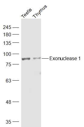 Sample:
Testis (Mouse) Lysate at 40 ug
Thymus (Mouse) Lysate at 40 ug
Primary: Anti-Exonuclease 1 (SL13119R) at 1/1000 dilution
Secondary: IRDye800CW Goat Anti-Rabbit IgG at 1/20000 dilution
Predicted band size: 94 kD
Observed band size: 90 kD
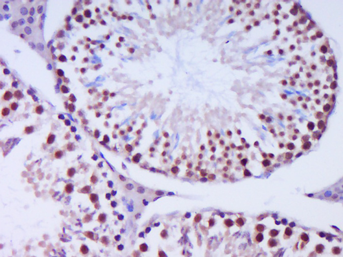 Paraformaldehyde-fixed, paraffin embedded (Mouse testis); Antigen retrieval by boiling in sodium citrate buffer (pH6.0) for 15min; Block endogenous peroxidase by 3% hydrogen peroxide for 20 minutes; Blocking buffer (normal goat serum) at 37°C for 30min; Antibody incubation with (Exonuclease 1) Polyclonal Antibody, Unconjugated (SL13119R) at 1:400 overnight at 4°C, followed by operating according to SP Kit(Rabbit) (sp-0023) instructionsand DAB staining.
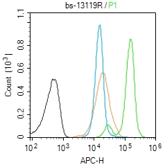 Blank control (Black line):Molt4 (Black).
Primary Antibody (green line): Rabbit Anti-Exonuclease 1 antibody (SL13119R)
Dilution: 1μg /10^6 cells;
Isotype Control Antibody (orange line): Rabbit IgG .
Secondary Antibody (white blue line): Goat anti-rabbit IgG-AF647
Dilution: 1μg /test.
Protocol
The cells were fixed with 4% PFA (10min at room temperature)and then permeabilized with 90% ice-cold methanol for 20 min at room temperature. The cells were then incubated in 5%BSA to block non-specific protein-protein interactions for 30 min at room temperature .Cells stained with Primary Antibody for 30 min at room temperature. The secondary antibody used for 40 min at room temperature. Acquisition of 20,000 events was performed.
Cartpieces
Totalgoods,subtotals:¥Checkout
Bought notes(bought amounts latest0)
No one bought this product
User Comment(Total0User Comment Num)
- No comment


 +86 571 56623320
 +86 18668110335

