
Rabbit Anti-PARK7/DJ1 antibody
PARK7; PARK-7; Parkinson disease protein 7; Park7 protein; CAP1; DJ-1; DJ 1; SP22; Protein DJ-1; Oncogene DJ1; FLJ27376; Park 7; Parkinson disease (autosomal recessive early onset) 7; RNA binding protein regulatory subunit; RS; SP22; CAP1_HUMAN.
View History [Clear]
Details
Product Name PARK7/DJ1 Chinese Name CAP1抗体 Alias PARK7; PARK-7; Parkinson disease protein 7; Park7 protein; CAP1; DJ-1; DJ 1; SP22; Protein DJ-1; Oncogene DJ1; FLJ27376; Park 7; Parkinson disease (autosomal recessive early onset) 7; RNA binding protein regulatory subunit; RS; SP22; CAP1_HUMAN. Research Area Tumour immunology Neurobiology Signal transduction Kinases and Phosphatases Epigenetics G protein signal Immunogen Species Rabbit Clonality Polyclonal React Species Human, Mouse, Rat, (predicted: Pig, Cow, Horse, ) Applications WB=1:500-2000 ELISA=1:5000-10000 IHC-P=1:100-500 IHC-F=1:100-500 Flow-Cyt=0.2μg/Test IF=1:100-500 (Paraffin sections need antigen repair)
not yet tested in other applications.
optimal dilutions/concentrations should be determined by the end user.Theoretical molecular weight 20kDa Cellular localization The nucleus cytoplasmic Form Liquid Concentration 1mg/ml immunogen KLH conjugated synthetic peptide derived from human CAP1: 101-189/189 Lsotype IgG Purification affinity purified by Protein A Buffer Solution 0.01M TBS(pH7.4) with 1% BSA, 0.03% Proclin300 and 50% Glycerol. Storage Shipped at 4℃. Store at -20 °C for one year. Avoid repeated freeze/thaw cycles. Attention This product as supplied is intended for research use only, not for use in human, therapeutic or diagnostic applications. PubMed PubMed Product Detail PARK7/DJ1 is a ubiquitously expressed protein involved in various cellular processes including cell proliferation, RNA-binding, and oxidative stress. The protein has been found to colocalize within a subset of pathologic tau inclusions in a diverse group of neurodegenerative disorders known as tauopathies (Rizzu et al. 2004). Defects in PARK7/DJ1 are the cause of autosomal recessive early-onset Parkinson's disease 7 (PARK7). Parkinson's disease (PD) is a complex, multifactorial disorder that typically manifests after the age of 50 years. The disease is characterized by bradykinesia, resting tremor, muscular rigidity and postural instability. The pathology involves the loss of dopaminergic neurons in the substantia nigra and the presence of Lewy bodies (intraneuronal accumulations of aggregated proteins), in surviving neurons in various areas of the brain. PARK7 is characterized by onset before 40 years and slow progression. It has also been suggested that PARK7/DJ1 is a mitogen dependent oncogene product involved in Ras related signal transduction pathways.
Function:
Protects cells against oxidative stress and cell death. Plays a role in regulating expression or stability of the mitochondrial uncoupling proteins SLC25A14 and SLC25A27 in dopaminergic neurons of the substantia nigra pars compacta and attenuates the oxidative stress induced by calcium entry into the neurons via L-type channels during pacemaking. Eliminates hydrogen peroxide and protects cells against hydrogen peroxide-induced cell death. May act as an atypical peroxiredoxin-like peroxidase that scavenges hydrogen peroxide. Following removal of a C-terminal peptide, displays protease activity and enhanced cytoprotective action against oxidative stress-induced apoptosis. Stabilizes NFE2L2 by preventing its association with KEAP1 and its subsequent ubiquitination. Binds to OTUD7B and inhibits its deubiquitinating activity. Enhances RELA nuclear translocation. Binds to a number of mRNAs containing multiple copies of GG or CC motifs and partially inhibits their translation but dissociates following oxidative stress. Required for correct mitochondrial morphology and function and for autophagy of dysfunctional mitochondria. Regulates astrocyte inflammatory responses. Acts as a positive regulator of androgen receptor-dependent transcription. Prevents aggregation of SNCA. Plays a role in fertilization. Has no proteolytic activity. Has cell-growth promoting activity and transforming activity. May function as a redox-sensitive chaperone.
Subcellular Location:
Cytoplasm. Nucleus. Mitochondrion. Under normal conditions, located predominantly in the cytoplasm and, to a lesser extent, in the nucleus and mitochondrion. Translocates to the mitochondrion and subsequently to the nucleus in response to oxidative stress and exerts an increased cytoprotective effect against oxidative damage. Detected in tau inclusions in brains from neurodegenerative disease patients.
Tissue Specificity:
Highly expressed in pancreas, kidney, skeletal muscle, liver, testis and heart. Detected at slightly lower levels in placenta and brain. Detected in astrocytes, Sertoli cells, spermatogonia, spermatids and spermatozoa.
Post-translational modifications:
Sumoylated on Lys-130 by PIAS2 or PIAS4; which is enhanced after ultraviolet irradiation and essential for cell-growth promoting activity and transforming activity.
DISEASE:
Defects in PARK7 are the cause of Parkinson disease type 7 (PARK7) [MIM:606324]. A neurodegenerative disorder characterized by resting tremor, postural tremor, bradykinesia, muscular rigidity, anxiety and psychotic episodes. PARK7 has onset before 40 years, slow progression and initial good response to levodopa. Some patients may show traits reminiscent of amyotrophic lateral sclerosis-parkinsonism/dementia complex (Guam disease).
Similarity:
Belongs to the peptidase C56 family.
SWISS:
Q99497
Gene ID:
11315
Database links:Entrez Gene: 11315 Human
Entrez Gene: 57320 Mouse
Omim: 602533 Human
SwissProt: Q99497 Human
SwissProt: Q99LX0 Mouse
Unigene: 419640 Human
Unigene: 277349 Mouse
Unigene: 30105 Rat
DJ-1蛋白是近年来新发现的一种Tumour蛋白,DJ-1能有效保护细胞抵抗氧化应激, 该蛋白早期主要用于Tumour方面的研究,近年来科研人员经研究认为:DJ-1蛋白与神经退化及损伤有一定的关联。Product Picture 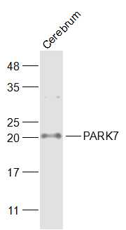 Sample:
Cerebrum (Mouse) Lysate at 40 ug
Primary: Anti-PARK7 (SL1306R) at 1/1000 dilution
Secondary: IRDye800CW Goat Anti-Rabbit IgG at 1/20000 dilution
Predicted band size: 20 kD
Observed band size: 20 kD
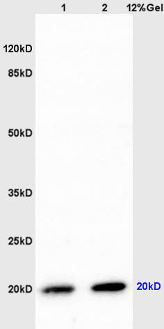 Sample:
Lane1:Brain(Rat) lysate at 30ug;
Lane2:Brain(Rat) lysate at 30ug;
Primary: Anti-CAP1/PARK7 (SL1306R) at 1:200 dilution;
Secondary: Alkaline phosphatase conjugated Goat Anti-Rabbit IgG(SL0295G-AP) at 1: 3000 dilution;
Predicted band size : 20kD
Observed band size : 20kD
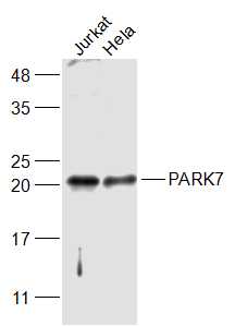 Sample:
Jurkat(Human) Cell Lysate at 30 ug
Hela(Human) Cell Lysate at 30 ug
Primary: Anti-PARK7 (SL1306R) at 1/1000 dilution
Secondary: IRDye800CW Goat Anti-Rabbit IgG at 1/20000 dilution
Predicted band size: 20 kD
Observed band size: 20 kD
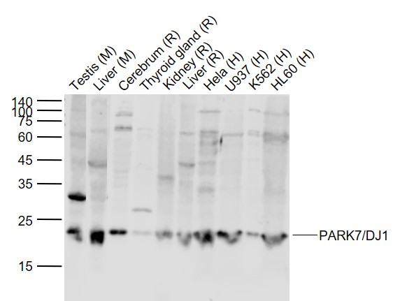 Sample:
Lane 1: Testis (Mouse) Lysate at 40 ug
Lane 2: Liver (Mouse) Lysate at 40 ug
Lane 3: Cerebrum (Rat) Lysate at 40 ug
Lane 4: Thyroid gland Rat) Lysate at 40 ug
Lane 5: Kidney (Rat) Lysate at 40 ug
Lane 6: Liver (Rat) Lysate at 40 ug
Lane 7: Hela (Human) Cell Lysate at 30 ug
Lane 8: U937 (Human) Cell Lysate at 30 ug
Lane 9: K562 (Human) Cell Lysate at 30 ug
Lane 10: HL60 (Human) Cell Lysate at 30 ug
Primary:
Anti-PARK7/DJ1 (SL1306R) at 1/1000 dilution
Secondary: IRDye800CW Goat Anti-Rabbit IgG at 1/20000 dilution
Predicted band size: 22 kD
Observed band size: 22 kD
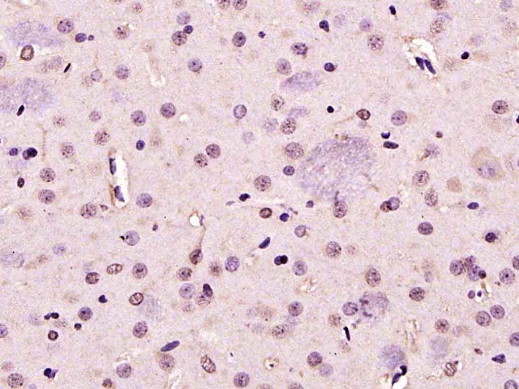 Paraformaldehyde-fixed, paraffin embedded (rat brain); Antigen retrieval by boiling in sodium citrate buffer (pH6.0) for 15min; Block endogenous peroxidase by 3% hydrogen peroxide for 20 minutes; Blocking buffer (normal goat serum) at 37°C for 30min; Antibody incubation with (PARK7) Polyclonal Antibody, Unconjugated (SL1306R ) at 1:200 overnight at 4°C, followed by operating according to SP Kit(Rabbit) (sp-0023) instructionsand DAB staining.
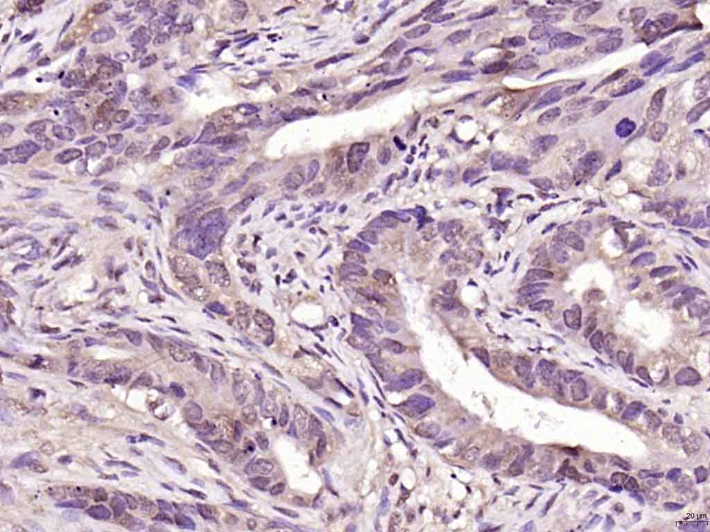 Paraformaldehyde-fixed, paraffin embedded (human colon carcinoma); Antigen retrieval by boiling in sodium citrate buffer (pH6.0) for 15min; Block endogenous peroxidase by 3% hydrogen peroxide for 20 minutes; Blocking buffer (normal goat serum) at 37°C for 30min; Antibody incubation with (PARK7) Polyclonal Antibody, Unconjugated (SL1306R ) at 1:200 overnight at 4°C, followed by operating according to SP Kit(Rabbit) (sp-0023) instructionsand DAB staining.
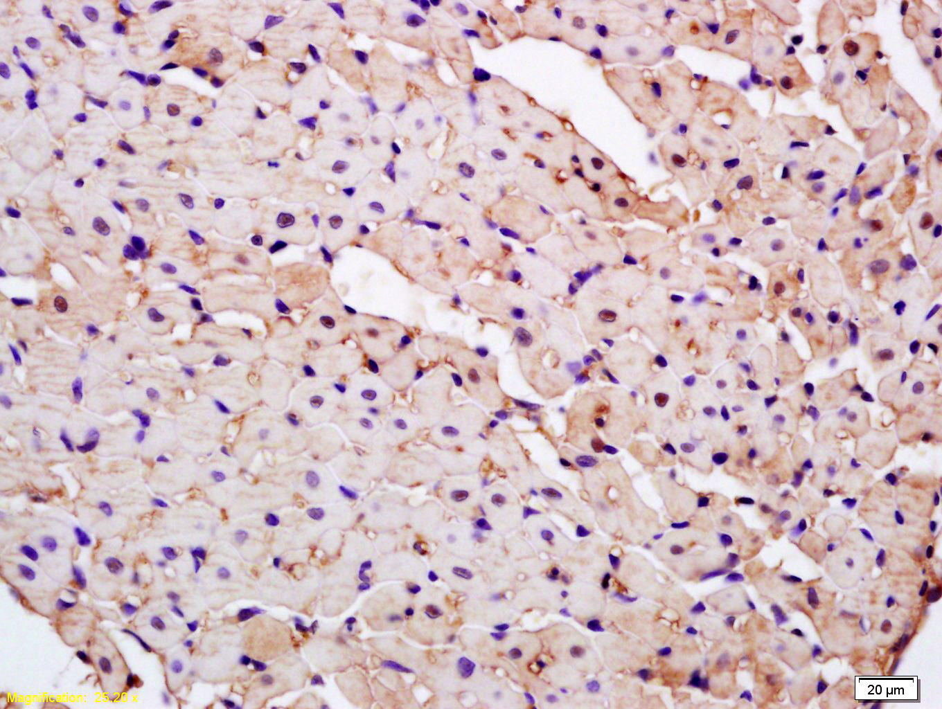 Tissue/cell: rat kidney tissue; 4% Paraformaldehyde-fixed and paraffin-embedded;
Antigen retrieval: citrate buffer ( 0.01M, pH 6.0 ), Boiling bathing for 15min; Block endogenous peroxidase by 3% Hydrogen peroxide for 30min; Blocking buffer (normal goat serum,C-0005) at 37℃ for 20 min;
Incubation: Anti-CAP1/PARK7 Polyclonal Antibody, Unconjugated(SL1306R) 1:200, overnight at 4°C, followed by conjugation to the secondary antibody(SP-0023) and DAB(C-0010) staining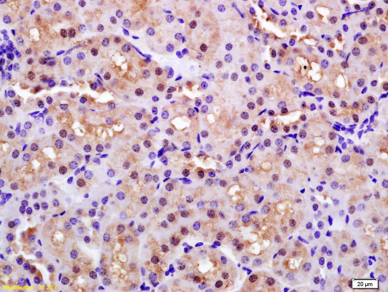
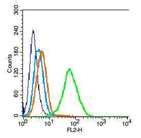 Blank control: 293T cells(blue).
Primary Antibody: Rabbit Anti-PARK7/CAP1 antibody(SL1306R), Dilution: 0.2μg in 100 μL 1X PBS containing 0.5% BSA;
Isotype Control Antibody: Rabbit IgG (orange) ,used under the same conditions.
Secondary Antibody: Goat anti-rabbit IgG-PE(white blue), Dilution: 1:200 in 1 X PBS containing 0.5% BSA.
Protocol
Primary antibody (SL1306R, 0.2μg /1x10^6 cells) were incubated for 30 min on the ice, followed by 1 X PBS containing 0.5% BSA + 1 0% goat serum (15 min) to block non-specific protein-protein interactions. Then the Goat Anti-rabbit IgG/PE antibody was added into the blocking buffer mentioned above to react with the primary antibody at 1/200 dilution for 30 min on ice. Acquisition of 20,000 events was performed. The blue histogram is unstained cells (Hepg2 cells)
concentration 1:50
The Wathet Blue histogram is cells stained with secondary antibody alone.
The Orange histogram is cells stained with rabbit IgG isotype control antibody plus secondary antibody.
The green histogram is cells stained with Rabbit Anti-PARK7/CAP1 antibody (SL1306R)plus secondary antibody.
Cartpieces
Totalgoods,subtotals:¥Checkout
Bought notes(bought amounts latest0)
No one bought this product
User Comment(Total0User Comment Num)
- No comment


 +86 571 56623320
 +86 18668110335

