
Rabbit Anti-CCR7 antibody
CCR7_HUMAN; BLR 2; BLR2; C C chemokine receptor type 7; C C CKR 7; CC chemokine receptor 7; CC chemokine receptor type 7; CC CKR 7; CCCKR7; CCR 7; CD 197; CD197; CD197 antigen; CDW197; Chemokine C C motif receptor 7; Chemokine C C receptor 7; Chemokine re
View History [Clear]
Details
Product Name CCR7 Chinese Name 细胞表面Chemokine受体7抗体 Alias CCR7_HUMAN; BLR 2; BLR2; C C chemokine receptor type 7; C C CKR 7; CC chemokine receptor 7; CC chemokine receptor type 7; CC CKR 7; CCCKR7; CCR 7; CD 197; CD197; CD197 antigen; CDW197; Chemokine C C motif receptor 7; Chemokine C C receptor 7; Chemokine receptor 7-like protein; EBI 1; EBI1; Ebi1h; EBV Induced G Protein Coupled Receptor 1; Epstein Barr virus induced G protein coupled receptor; Epstein Barr virus induced gene 1; EVI 1; EVI1; Lymphocyte Specific G Protein Coupled Peptide Receptor; MGC108519; MIP 3 beta receptor; MIP3 Beta Receptor. literatures Research Area immunology Signal transduction G protein-coupled receptor G protein signal Immunogen Species Rabbit Clonality Polyclonal React Species Human, Mouse, Rat, (predicted: Dog, ) Applications WB=1:500-2000 ELISA=1:5000-10000 IHC-P=1:100-500 IHC-F=1:100-500 Flow-Cyt=1μg/Test ICC=1:100 IF=1:100-500 (Paraffin sections need antigen repair)
not yet tested in other applications.
optimal dilutions/concentrations should be determined by the end user.Theoretical molecular weight 42kDa Cellular localization The cell membrane Form Liquid Concentration 1mg/ml immunogen KLH conjugated synthetic peptide derived from human CCR7: 25-59/379 <Extracellular> Lsotype IgG Purification affinity purified by Protein A Buffer Solution 0.01M TBS(pH7.4) with 1% BSA, 0.03% Proclin300 and 50% Glycerol. Storage Shipped at 4℃. Store at -20 °C for one year. Avoid repeated freeze/thaw cycles. Attention This product as supplied is intended for research use only, not for use in human, therapeutic or diagnostic applications. PubMed PubMed Product Detail The protein encoded by this gene is a member of the G protein-coupled receptor family. This receptor was identified as a gene induced by the Epstein-Barr virus (EBV), and is thought to be a mediator of EBV effects on B lymphocytes. This receptor is expressed in various lymphoid tissues and activates B and T lymphocytes. It has been shown to control the migration of memory T cells to inflamed tissues, as well as stimulate dendritic cell maturation. The chemokine (C-C motif) ligand 19 (CCL19/ECL) has been reported to be a specific ligand of this receptor. [provided by RefSeq, Jul 2008]
Function:
Receptor for the MIP-3-beta chemokine. Probable mediator of EBV effects on B-lymphocytes or of normal lymphocyte functions.
Subcellular Location:
Cell membrane; Multi-pass membrane protein.
Tissue Specificity:
Expressed in various lymphoid tissues and activated B- and T-lymphocytes, strongly up-regulated in B-cells infected with Epstein-Barr virus and T-cells infected with herpesvirus 6 or 7.
Similarity:
Belongs to the G-protein coupled receptor 1 family.
SWISS:
P47774
Gene ID:
1236
Database links:Entrez Gene: 1236 Human
Entrez Gene: 12775 Mouse
Omim: 600242 Human
SwissProt: P32248 Human
SwissProt: P47774 Mouse
Unigene: 370036 Human
Unigene: 2932 Mouse
Unigene: 229736 Rat
Chemokine是当今cell factorResearch Area的热点之一,它参与多种免疫及炎症反应,在感染、Tumour的生长与转移、组织修复及创伤愈合等病理生理过程中发挥重要作用.CCR7在Tumour的生长与转移方面起到一定的作用.Product Picture 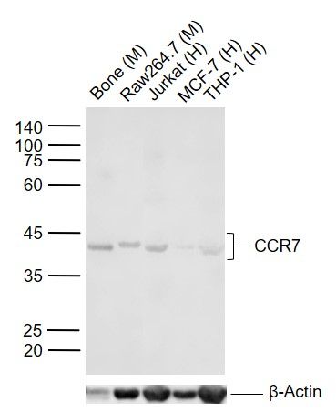 Sample:
Lane 1: Mouse Bone tissue lysates
Lane 2: Mouse Raw264.7 cell lysates
Lane 3: Human Jurkat cell lysates
Lane 4: Human MCF-7 cell lysates
Lane 5: Human THP-1 cell lysates
Primary: Anti-CCR7 (SL1305R) at 1/1000 dilution
Secondary: IRDye800CW Goat Anti-Rabbit IgG at 1/20000 dilution
Predicted band size: 42 kDa
Observed band size: 42 kDa
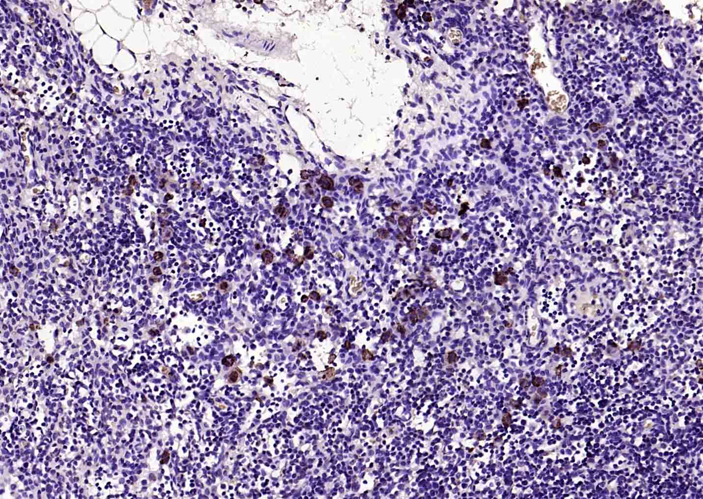 Paraformaldehyde-fixed, paraffin embedded (RAT lymphoid); Antigen retrieval by boiling in sodium citrate buffer (pH6.0) for 15min; Block endogenous peroxidase by 3% hydrogen peroxide for 20 minutes; Blocking buffer (normal goat serum) at 37°C for 30min; Antibody incubation with (CCR7) Polyclonal Antibody, Unconjugated (SL1305R) at 1:200 overnight at 4°C, followed by operating according to SP Kit(Rabbit) (sp-0023) instructionsand DAB staining.
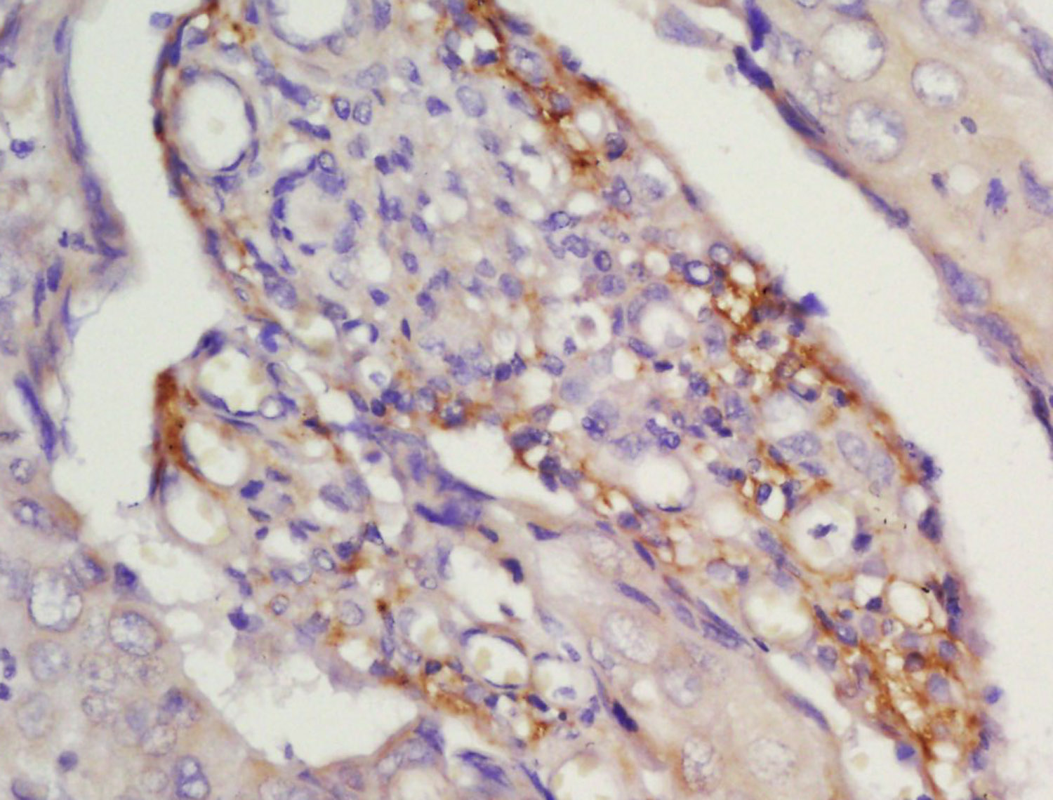 Tissue/cell: human laryngocarcinoma; 4% Paraformaldehyde-fixed and paraffin-embedded;
Antigen retrieval: citrate buffer ( 0.01M, pH 6.0 ), Boiling bathing for 15min; Block endogenous peroxidase by 3% Hydrogen peroxide for 30min; Blocking buffer (normal goat serum,C-0005) at 37℃ for 20 min;
Incubation: Anti-CCR7 Polyclonal Antibody, Unconjugated(SL1305R) 1:200, overnight at 4°C, followed by conjugation to the secondary antibody(SP-0023) and DAB(C-0010) staining
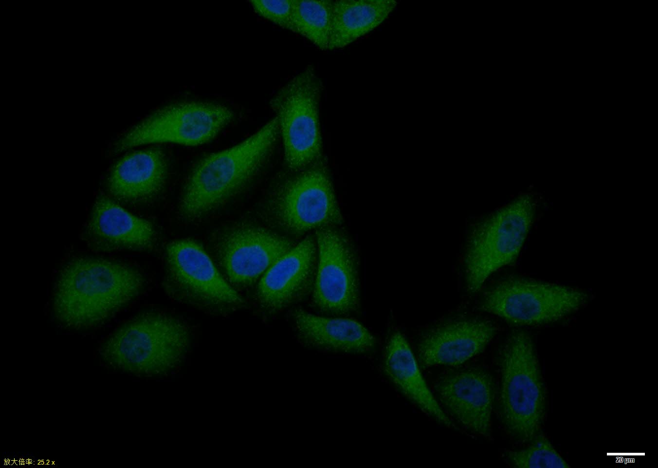 Hela cell; 4% Paraformaldehyde-fixed; Triton X-100 at room temperature for 20 min; Blocking buffer (normal goat serum, C-0005) at 37°C for 20 min; Antibody incubation with (CCR7) polyclonal Antibody, Unconjugated (SL1305R) 1:100, 90 minutes at 37°C; followed by a conjugated Goat Anti-Rabbit IgG antibody at 37°C for 90 minutes, DAPI (blue, C02-04002) was used to stain the cell nuclei.
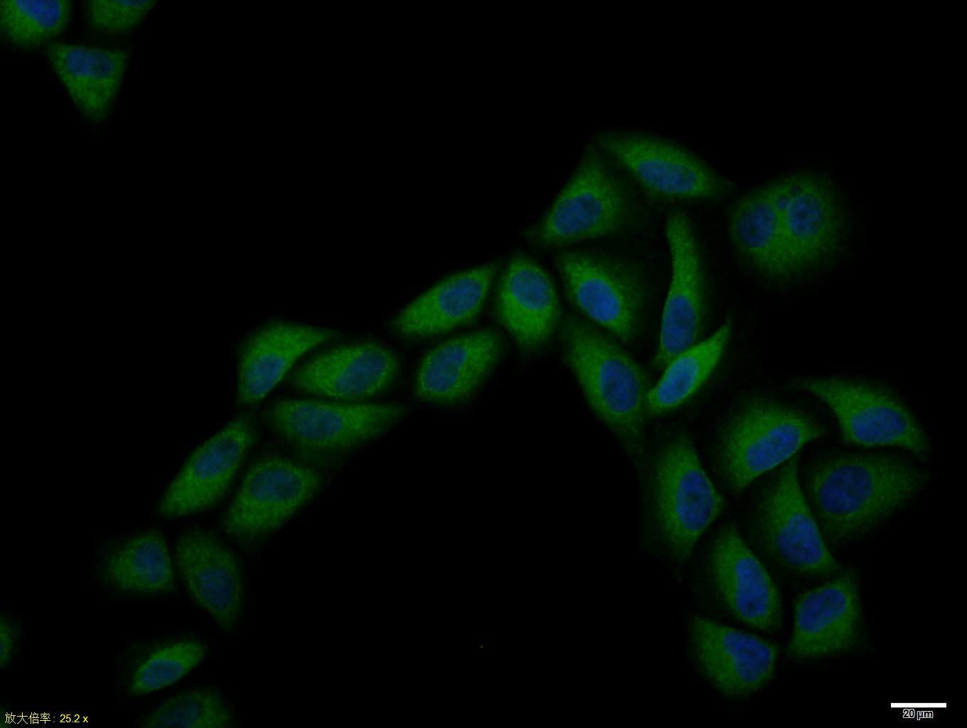 Hela cell; 4% Paraformaldehyde-fixed; Triton X-100 at room temperature for 20 min; Blocking buffer (normal goat serum, C-0005) at 37°C for 20 min; Antibody incubation with (CCR7) polyclonal Antibody, Unconjugated (SL1305R) 1:100, 90 minutes at 37°C; followed by a conjugated Goat Anti-Rabbit IgG antibody at 37°C for 90 minutes, DAPI (blue, C02-04002) was used to stain the cell nuclei.
 Tissue/cell: human gastric tissue;4% Paraformaldehyde-fixed and paraffin-embedded;
Antigen retrieval: citrate buffer ( 0.01M, pH 6.0 ), Boiling bathing for 15min; Blocking buffer (normal goat serum,C-0005) at 37℃ for 20 min;
Incubation: Anti-CCR7 Polyclonal Antibody, Unconjugated(SL1305R) 1:200, overnight at 4°C; The secondary antibody was Goat Anti-Rabbit IgG, FITC conjugated(SL0295G-FITC)used at 1:200 dilution for 40 minutes at 37°C. DAPI(5ug/ml,blue,C-0033) was used to stain the cell nuclei
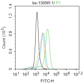 Blank control:THP-1.
Primary Antibody (green line): Rabbit Anti-CCR7 antibody (SL1305R)
Dilution: 1μg /10^6 cells;
Isotype Control Antibody (orange line): Rabbit IgG .
Secondary Antibody : Goat anti-rabbit IgG-FITC
Dilution: 0.5μg /test.
Protocol
The cells were fixed with 4% PFA (10min at room temperature)and then permeabilized with 0.1% PBST for 20 min at room temperature.The cells were then incubated in 5%BSA to block non-specific protein-protein interactions for 30 min at room temperature .Cells stained with Primary Antibody for 30 min at room temperature. The secondary antibody used for 40 min at room temperature. Acquisition of 20,000 events was performed.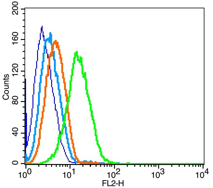 Blank control: Raji(blue).
Primary Antibody:Rabbit Anti-CCR7 antibody(SL1305R), Dilution: 1μg in 100 μL 1X PBS containing 0.5% BSA;
Isotype Control Antibody: Rabbit IgG(orange) ,used under the same conditions );
Secondary Antibody: Goat anti-rabbit IgG-PE(white blue), Dilution: 1:200 in 1 X PBS containing 0.5% BSA.
Protocol
The cells were fixed with 2% paraformaldehyde (10 min) . Primary antibody (SL1305R, 1μg /1x10^6 cells) were incubated for 30 min on the ice, followed by 1 X PBS containing 0.5% BSA + 10% goat serum (15 min) to block non-specific protein-protein interactions. Then the Goat Anti-rabbit IgG/PE antibody was added into the blocking buffer mentioned above to react with the primary antibody at 1/200 dilution for 30 min on ice. Acquisition of 20,000 events was performed.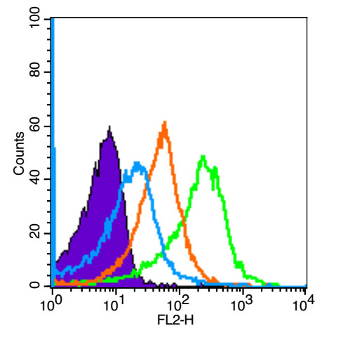 Blank control (Black line): Mouse spleen(Black). Primary Antibody (green line): Rabbit Anti-CD4 antibody (SL1305R-PE) Dilution: 3μg /10^6 cells; Isotype Control Antibody (orange line): Rabbit IgG-PE. Secondary Antibody (white blue line): Goat anti-rabbit IgG-PE
Dilution: 1μg /test.
Protocol The cells were fixed with 4% PFA (10min at room temperature)and then permeabilized with 90% ice-cold methanol for 20 min at room temperature. The cells were then incubated in 5%BSA to block non-specific protein-protein interactions for 30 min at room temperature .Cells stained with Primary Antibody for 30 min at room temperature.The secondary antibody used for 40 min at room temperature. Acquisition of 20,000 events was performed.
Cartpieces
Totalgoods,subtotals:¥Checkout
Bought notes(bought amounts latest0)
No one bought this product
User Comment(Total0User Comment Num)
- No comment


 +86 571 56623320
 +86 18668110335

