
Rabbit Anti-ICAM2 antibody
CD102; Intercellular adhesion molecule 2; CD102 antigen; ICAM 2; ICAM-2; Intercellular adhesion molecule 2 precursor; Ly60; ICAM2_HUMAN;
View History [Clear]
Details
Product Name ICAM2 Chinese Name 细胞间粘附分子-2(CD102)抗体 Alias CD102; Intercellular adhesion molecule 2; CD102 antigen; ICAM 2; ICAM-2; Intercellular adhesion molecule 2 precursor; Ly60; ICAM2_HUMAN; Research Area Tumour Cell adhesion molecule Cell Surface Molecule Immunogen Species Rabbit Clonality Polyclonal React Species Human, (predicted: Mouse, Rat, ) Applications WB=1:500-2000 ELISA=1:5000-10000 Flow-Cyt=1μg/Test
not yet tested in other applications.
optimal dilutions/concentrations should be determined by the end user.Theoretical molecular weight 28kDa Detection molecular weight 55 kDa Cellular localization The cell membrane Form Liquid Concentration 1mg/ml immunogen KLH conjugated synthetic peptide derived from human ICAM-2: 175-277/277 Lsotype IgG Purification affinity purified by Protein A Buffer Solution 0.01M TBS(pH7.4) with 1% BSA, 0.03% Proclin300 and 50% Glycerol. Storage Shipped at 4℃. Store at -20 °C for one year. Avoid repeated freeze/thaw cycles. Attention This product as supplied is intended for research use only, not for use in human, therapeutic or diagnostic applications. PubMed PubMed Product Detail The protein encoded by this gene is a member of the intercellular adhesion molecule (ICAM) family. All ICAM proteins are type I transmembrane glycoproteins, contain 2-9 immunoglobulin-like C2-type domains, and bind to the leukocyte adhesion LFA-1 protein. This protein may play a role in lymphocyte recirculation by blocking LFA-1-dependent cell adhesion. It mediates adhesive interactions important for antigen-specific immune response, NK-cell mediated clearance, lymphocyte recirculation, and other cellular interactions important for immune response and surveillance. Several transcript variants encoding the same protein have been found for this gene. [provided by RefSeq, Jul 2008]
Function:
ICAM proteins are ligands for the leukocyte adhesion protein LFA-1 (integrin alpha-L/beta-2). ICAM2 may play a role in lymphocyte recirculation by blocking LFA-1-dependent cell adhesion. It mediates adhesive interactions important for antigen-specific immune response, NK-cell mediated clearance, lymphocyte recirculation, and other cellular interactions important for immune response and surveillance.
Subcellular Location:
Membrane; Single-pass type I membrane protein.
Tissue Specificity:
Expressed in endothelial cells and leukocytes. High levels found in lung.
Similarity:
Belongs to the immunoglobulin superfamily. ICAM family.
Contains 2 Ig-like C2-type (immunoglobulin-like) domains.
SWISS:
P13598
Gene ID:
3384
Database links:Entrez Gene: 3384 Human
Entrez Gene: 15896 Mouse
Omim: 146630 Human
SwissProt: P13598 Human
SwissProt: P35330 Mouse
ICAM-2也是免疫蛋白超家族粘附分子之一.Product Picture 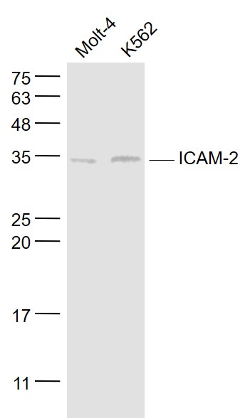 Sample:
Molt-4(Human) Cell Lysate at 30 ug
K562(Human) Cell Lysate at 30 ug
Primary: Anti- ICAM-2/CD102 (SL1258R) at 1/1000 dilution
Secondary: IRDye800CW Goat Anti-Rabbit IgG at 1/20000 dilution
Predicted band size: 28 kD
Observed band size: 31 kD
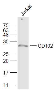 Sample:
Jurkat(Human) Cell Lysate at 30 ug
Primary: Anti-CD102 (SL1258R) at 1/1000 dilution
Secondary: IRDye800CW Goat Anti-Rabbit IgG at 1/20000 dilution
Predicted band size: 28 kD
Observed band size: 28 kD
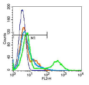 Blank control: Jurkat cells(blue).
Primary Antibody:Rabbit Anti-CD102 antibody(SL1258R), Dilution: 1μg in 100 μL 1X PBS containing 0.5% BSA;
Isotype Control Antibody: Rabbit IgG(orange) ,used under the same conditions );
Secondary Antibody: Goat anti-rabbit IgG-PE(white blue), Dilution: 1:200 in 1 X PBS containing 0.5% BSA.
Protocol
The cells were fixed with 2% paraformaldehyde (10 min) . Primary antibody (SL1258R, 1μg /1x10^6 cells) were incubated for 30 min on the ice, followed by 1 X PBS containing 0.5% BSA + 1 0% goat serum (15 min) to block non-specific protein-protein interactions. Then the Goat Anti-rabbit IgG/PE antibody was added into the blocking buffer mentioned above to react with the primary antibody at 1/200 dilution for 30 min on ice. Acquisition of 20,000 events was performed.
Cartpieces
Totalgoods,subtotals:¥Checkout
Bought notes(bought amounts latest0)
No one bought this product
User Comment(Total0User Comment Num)
- No comment


 +86 571 56623320
 +86 18668110335

