
Rabbit Anti-phospho-APC (Ser2054)antibody
APC (phospho S2054); p-APC (phospho S2054); Adenomatous Polyposis Coli; APC; CC1; DP2; DP2.5; DP3; FAP; FPC; GS; Protein APC; APC_HUMAN.
View History [Clear]
Details
Product Name phospho-APC (Ser2054) Chinese Name 磷酸化腺瘤样息肉抗体 Alias APC (phospho S2054); p-APC (phospho S2054); Adenomatous Polyposis Coli; APC; CC1; DP2; DP2.5; DP3; FAP; FPC; GS; Protein APC; APC_HUMAN. Product Type Phosphorylated anti Research Area Tumour Cell biology Developmental biology Neurobiology Signal transduction Stem cells Epigenetics Immunogen Species Rabbit Clonality Polyclonal React Species Human, Mouse, Rat, (predicted: Dog, Pig, Cow, Rabbit, Sheep, ) Applications IHC-P=1:100-500 IHC-F=1:100-500 IF=1:100-500 (Paraffin sections need antigen repair)
not yet tested in other applications.
optimal dilutions/concentrations should be determined by the end user.Theoretical molecular weight 312kDa Cellular localization cytoplasmic The cell membrane Form Liquid Concentration 1mg/ml immunogen KLH conjugated synthesised phosphopeptide derived from human APC around the phosphorylation site of Ser2054: KP(p-S)RL Lsotype IgG Purification affinity purified by Protein A Buffer Solution 0.01M TBS(pH7.4) with 1% BSA, 0.03% Proclin300 and 50% Glycerol. Storage Shipped at 4℃. Store at -20 °C for one year. Avoid repeated freeze/thaw cycles. Attention This product as supplied is intended for research use only, not for use in human, therapeutic or diagnostic applications. PubMed PubMed Product Detail Tumor suppressor. Promotes rapid degradation of CTNNB1 and participates in Wnt signaling as a negative regulator. APC activity is correlated with its phosphorylation state. Activates the GEF activity of SPATA13 and ARHGEF4. Plays a role in hepatocyte growth factor (HGF)-induced cell migration. Required for MMP9 up-regulation via the JNK signaling pathway in colorectal tumor cells. Acts as a mediator of ERBB2-dependent stabilization of microtubules at the cell cortex. It is required for the localization of MACF1 to the cell membrane and this localization of MACF1 is critical for its function in microtubule stabilization.
Subcellular Location:
Cell junction > adherens junction. Cytoplasm > cytoskeleton. Cell projection > lamellipodium. Cell projection > ruffle membrane. Cytoplasm. Cell membrane. Associated with the microtubule network at the growing distal tip of microtubules. Accumulates in the lamellipodium and ruffle membrane in response to hepatocyte growth factor (HGF) treatment. The MEMO1-RHOA-DIAPH1 signaling pathway controls localization of the phosophorylated form to the cell membrane.
Tissue Specificity:
Expressed in a variety of tissues.
Post-translational modifications:
Phosphorylated by GSK3B.
Ubiquitinated, leading to its degradation by the proteasome. Ubiquitination is facilitated by Axin. Deubiquitinated by ZRANB1/TRABID.
DISEASE:
Defects in APC are a cause of familial adenomatous polyposis (FAP) [MIM:175100]; which includes also Gardner syndrome (GS). FAP and GS contribute to tumor development in patients with uninherited forms of colorectal cancer. FAP is characterized by adenomatous polyps of the colon and rectum, but also of upper gastrointestinal tract (ampullary, duodenal and gastric adenomas). This is a viciously premalignant disease with one or more polyps progressing through dysplasia to malignancy in untreated gene carriers with a median age at diagnosis of 40 years.
Defects in APC are a cause of hereditary desmoid disease (HDD) [MIM:135290]; also known as familial infiltrative fibromatosis (FIF). HDD is an autosomal dominant trait with 100% penetrance and possible variable expression among affected relatives. HDD patients show multifocal fibromatosis of the paraspinal muscles, breast, occiput, arms, lower ribs, abdominal wall, and mesentery. Desmoid tumors appears also as a complication of familial adenomatous polyposis.
Defects in APC are a cause of medulloblastoma (MDB) [MIM:155255]. MDB is a malignant, invasive embryonal tumor of the cerebellum with a preferential manifestation in children. Although the majority of medulloblastomas occur sporadically, some manifest within familial cancer syndromes such as Turcot syndrome and basal cell nevus syndrome (Gorlin syndrome). Defects in APC are a cause of mismatch repair cancer syndrome (MMRCS) [MIM:276300]; also known as Turcot syndrome or brain tumor-polyposis syndrome 1 (BTPS1). MMRCS is an autosomal dominant disorder characterized by malignant tumors of the brain associated with multiple colorectal adenomas. Skin features include sebaceous cysts, hyperpigmented and cafe au lait spots.
Defects in APC are a cause of gastric cancer (GASC) [MIM:613659]; also called gastric cancer intestinal or stomach cancer. Gastric cancer is a malignant disease which starts in the stomach, can spread to the esophagus or the small intestine, and can extend through the stomach wall to nearby lymph nodes and organs. It also can metastasize to other parts of the body. The term gastric cancer or gastric carcinoma refers to adenocarcinoma of the stomach that accounts for most of all gastric malignant tumors. Two main histologic types are recognized, diffuse type and intestinal type carcinomas. Diffuse tumors are poorly differentiated infiltrating lesions, resulting in thickening of the stomach. In contrast, intestinal tumors are usually exophytic, often ulcerating, and associated with intestinal metaplasia of the stomach, most often observed in sporadic disease.
Defects in APC are a cause of hepatocellular carcinoma (HCC) [MIM:114550]. This defect includes also the disease entity termed hepatoblastoma.
Similarity:
Belongs to the adenomatous polyposis coli (APC) family.
Contains 7 ARM repeats.
SWISS:
P25054
Gene ID:
324
Database links:Entrez Gene: 324 Human
Omim: 611731 Human
SwissProt: P25054 Human
Unigene: 158932 Human
Unigene: 88057 Rat
Product Picture 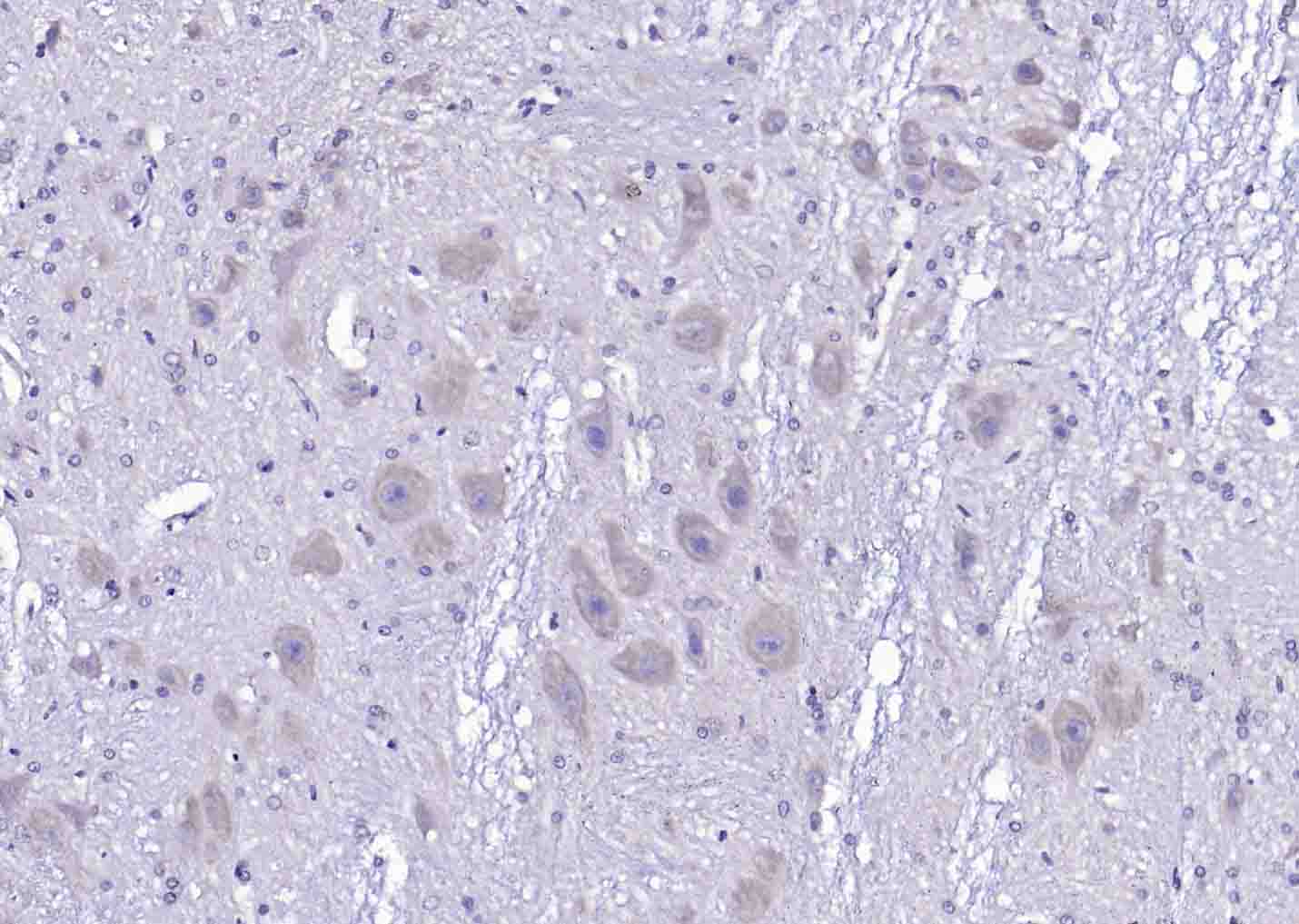 Paraformaldehyde-fixed, paraffin embedded (rat cerebellum); Antigen retrieval by boiling in sodium citrate buffer (pH6.0) for 15min; Block endogenous peroxidase by 3% hydrogen peroxide for 20 minutes; Blocking buffer (normal goat serum) at 37°C for 30min; Antibody incubation with (phospho-APC (Ser2054)) Polyclonal Antibody, Unconjugated (SL12481R) at 1:200 overnight at 4°C, followed by operating according to SP Kit(Rabbit) (sp-0023) instructionsand DAB staining.
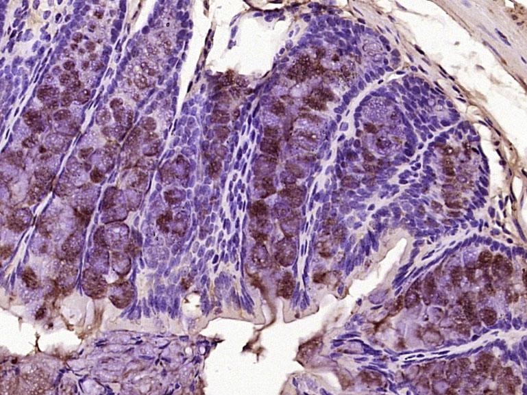 Paraformaldehyde-fixed, paraffin embedded (mouse colon tissue); Antigen retrieval by boiling in sodium citrate buffer (pH6.0) for 15min; Block endogenous peroxidase by 3% hydrogen peroxide for 20 minutes; Blocking buffer (normal goat serum) at 37°C for 30min; Antibody incubation with (APC (Ser2054)) Polyclonal Antibody, Unconjugated (SL12481R) at 1:400 overnight at 4°C, followed by operating according to SP Kit(Rabbit) (sp-0023) instructionsand DAB staining.
Cartpieces
Totalgoods,subtotals:¥Checkout
Bought notes(bought amounts latest0)
No one bought this product
User Comment(Total0User Comment Num)
- No comment


 +86 571 56623320
 +86 18668110335

