
Rabbit Anti-P-cadherin antibody
placental; Cadherin-3; P cadherin; Cadp; Placental cadherin; P-cadherin; AI385538; Pca; Cadherin 3 precursor; Cadherin 3 type 1; Calcium dependent adhesion protein placental; Cadherin; CDH3; CDHP; 7B4 antigen; Cadherin-1; Cadherin-2; Cadherin-4; Cadherin-
View History [Clear]
Details
Product Name P-cadherin Chinese Name P-钙粘附分子抗体 Alias placental; Cadherin-3; P cadherin; Cadp; Placental cadherin; P-cadherin; AI385538; Pca; Cadherin 3 precursor; Cadherin 3 type 1; Calcium dependent adhesion protein placental; Cadherin; CDH3; CDHP; 7B4 antigen; Cadherin-1; Cadherin-2; Cadherin-4; Cadherin-5; CAM 120/80; CD144; CD324; CD325; Cdh4; CDH5; CDHE; CDHN; CADH3_HUMAN; E-cadherin; Epithelial cadherin; N-cadherin; NCAD; Neural cadherin; P-cadherin; UVO; Uvomorulin; Vascular endothelial cadherin; CDH 3; CDH3; CDH3 protein; CDHP; HJMD; PCAD; Placental cadherin. literatures Research Area Cell biology immunology Cell adhesion molecule Immunogen Species Rabbit Clonality Polyclonal React Species Human, (predicted: Mouse, Rat, Chicken, Dog, Cow, Rabbit, Guinea Pig, ) Applications WB=1:500-2000 ELISA=1:5000-10000 IHC-P=1:100-500 IHC-F=1:100-500 Flow-Cyt=2μg/Test ICC=1:100-500 IF=1:100-500 (Paraffin sections need antigen repair)
not yet tested in other applications.
optimal dilutions/concentrations should be determined by the end user.Theoretical molecular weight 80kDa Cellular localization The cell membrane Form Liquid Concentration 1mg/ml immunogen KLH conjugated synthetic peptide derived from human P-cadherin: 625-725/829 <Cytoplasmic> Lsotype IgG Purification affinity purified by Protein A Buffer Solution 0.01M TBS(pH7.4) with 1% BSA, 0.03% Proclin300 and 50% Glycerol. Storage Shipped at 4℃. Store at -20 °C for one year. Avoid repeated freeze/thaw cycles. Attention This product as supplied is intended for research use only, not for use in human, therapeutic or diagnostic applications. PubMed PubMed Product Detail
cell-cell adhesion molecules. Cadherins are responsible for a whole range of processes including development, wound healing, cell-cell signaling, cell growth and differentiation. N-cadherin is found in many locations including cardiac adherins junctions, oral squamous epithelial cells, and breast epithelial cells. Studies have linked N-cadherin to cancer metastasis by showing the aggressive tumor cells had preferentially turned on N-cadherin as opposed to E- or P-cadherin.
Cadherins are calcium dependent cell adhesion proteins. They preferentially interact with themselves in a homophilic manner in connecting cells; cadherins may thus contribute to the sorting of heterogeneous cell types.
Function:
Cadherins are calcium dependent cell adhesion proteins. They preferentially interact with themselves in a homophilic manner in connecting cells; cadherins may thus contribute to the sorting of heterogeneous cell types.
Subunit:
Interacts with CDCP1.
Subcellular Location:
Cell membrane; Single-pass type I membrane protein.
Tissue Specificity:
Expressed in some normal epithelial tissues and in some carcinoma cell lines.
DISEASE:
Defects in CDH3 are the cause of hypotrichosis with juvenile macular dystrophy (HJMD) [MIM:601553]. HJMD is a rare autosomal recessive disorder characterized by early hair loss heralding severe degenerative changes of the retinal macula and culminating in blindness during the second to third decade of life.
Defects in CDH3 are the cause of ectodermal dysplasia with ectrodactyly and macular dystrophy (EEM) [MIM:225280]; also known as EEM syndrome, Albrectsen-Svendsen syndrome or Ohdo-Hirayama-Terawaki syndrome. Ectodermal dysplasia defines a heterogeneous group of disorders due to abnormal development of two or more ectodermal structures. EEM is an autosomal recessive condition characterized by features of ectodermal dysplasia such as sparse eyebrows and scalp hair, and selective tooth agenesis associated with macular dystrophy and ectrodactyly.
Similarity:
Contains 5 cadherin domains.
SWISS:
P22223
Gene ID:
1001
Database links:Entrez Gene: 1001 Human
Omim: 114021 Human
SwissProt: P22223 Human
Unigene: 191842 Human
Product Picture 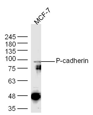 Sample:
MCF-7(Human) Cell Lysate at 30 ug
Primary: Anti-P-cadherin (SL1159R) at 1/300 dilution
Secondary: IRDye800CW Goat Anti-Rabbit IgG at 1/20000 dilution
Predicted band size: 80 kD
Observed band size: 90 kD
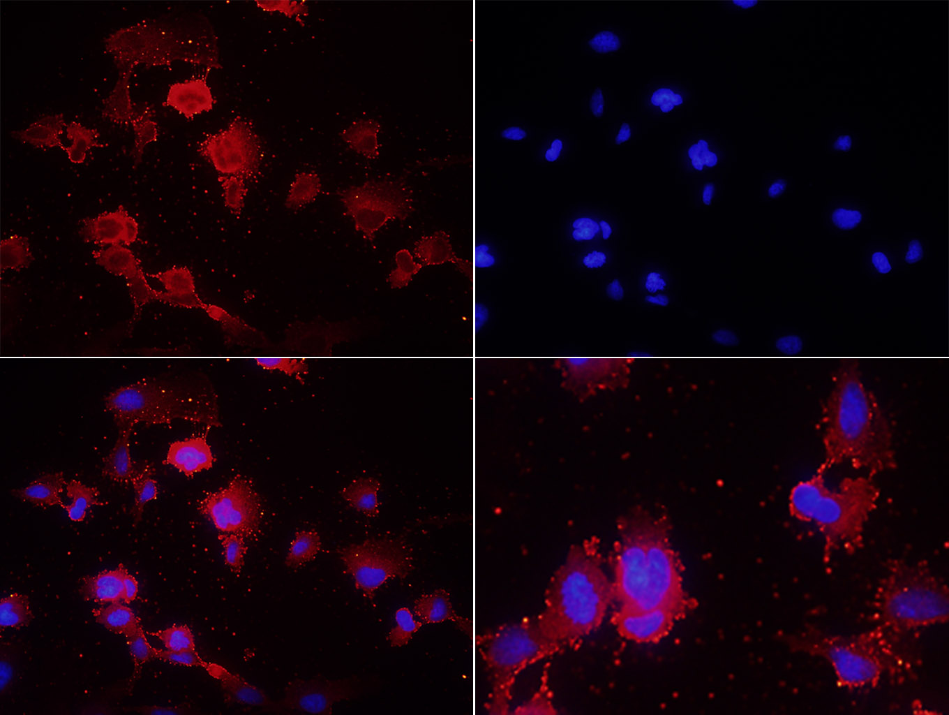 Tissue/cell: U251 cells;4% Paraformaldehyde-fixed;
Blocking buffer (normal goat serum,C-0005) at 37℃ for 20 min;
Incubation: Anti-P-cadherin Polyclonal Antibody, Unconjugated(SL1159R) 1:200, overnight at 4°C; The secondary antibody was Goat Anti-Rabbit IgG, Cy3 conjugated(SL0295G-Cy3)used at 1:200 dilution for 40 minutes at 37°C. DAPI(5ug/ml,blue,C-0033) was used to stain the cell nuclei
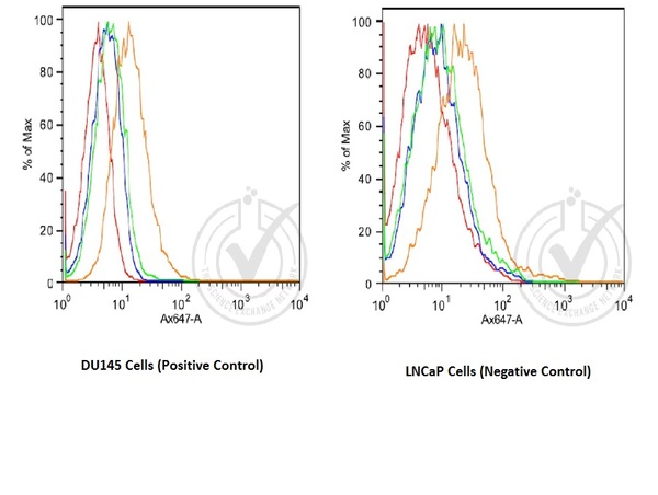 Image provided by Independent Validation (badge 029741). Histogram of human DU145 and human LNCaP cells stained with Rabbit Anti-P-cadherin Polyclonal Antibody (orange)(SL1159R at 1:100), isotype control antibody (green), secondary antibody only (blue) and unstained (red)
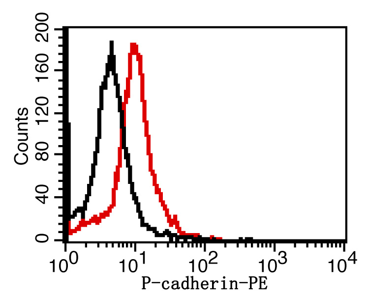 Overlay histogram showing Mouse SP2/0 stained with SL1159R-PE (red line). The cells were fixed with 1% paraformaldehyde (10 min). The cells were then incubated with the antibody(SL1159R-PE, 2ug/1x106cells) for 30 min at 22-25°C. Isotype control antibody (black line) was rabbit IgG (2ug/1x106cells) used under the same conditions. Acquisition of >5,000 events was performed.
Cartpieces
Totalgoods,subtotals:¥Checkout
Bought notes(bought amounts latest0)
No one bought this product
User Comment(Total0User Comment Num)
- No comment


 +86 571 56623320
 +86 18668110335

