
Rabbit Anti-PD-L1 antibody
Programmed cell death 1 ligand 1; CD274; B7 H; B7 H1; B7 homolog 1; B7-H1; B7H; B7H1; CD 274; CD274 antigen; CD274 molecule; MGC142294; MGC142296; OTTHUMP00000021029; PD L1; PD1L1_HUMAN; PDCD1 ligand 1; PDCD1L1; PDCD1LG1; PDL 1; PDL1; Programmed death lig
View History [Clear]
Details
Product Name PD-L1 Chinese Name 程序性死亡配体1(CD274)抗体 Alias Programmed cell death 1 ligand 1; CD274; B7 H; B7 H1; B7 homolog 1; B7-H1; B7H; B7H1; CD 274; CD274 antigen; CD274 molecule; MGC142294; MGC142296; OTTHUMP00000021029; PD L1; PD1L1_HUMAN; PDCD1 ligand 1; PDCD1L1; PDCD1LG1; PDL 1; PDL1; Programmed death ligand 1; RGD1566211. literatures Research Area Tumour immunology Cell Surface Molecule Cell type markers Immunogen Species Rabbit Clonality Polyclonal React Species Human, Mouse, Rat, (predicted: Pig, Cow, Horse, Sheep, ) Applications WB=1:500-2000 ELISA=1:5000-10000
not yet tested in other applications.
optimal dilutions/concentrations should be determined by the end user.Theoretical molecular weight 32kDa Cellular localization The cell membrane Form Liquid Concentration 1mg/ml immunogen KLH conjugated synthetic peptide derived from human CD274: 64-160/290 <Extracellular> Lsotype IgG Purification affinity purified by Protein A Buffer Solution 0.01M TBS(pH7.4) with 1% BSA, 0.03% Proclin300 and 50% Glycerol. Storage Shipped at 4℃. Store at -20 °C for one year. Avoid repeated freeze/thaw cycles. Attention This product as supplied is intended for research use only, not for use in human, therapeutic or diagnostic applications. PubMed PubMed Product Detail This gene encodes an immune inhibitory receptor ligand that is expressed by hematopoietic and non-hematopoietic cells, such as T cells and B cells and various types of tumor cells. The encoded protein is a type I transmembrane protein that has immunoglobulin V-like and C-like domains. Interaction of this ligand with its receptor inhibits T-cell activation and cytokine production. During infection or inflammation of normal tissue, this interaction is important for preventing autoimmunity by maintaining homeostasis of the immune response. In tumor microenvironments, this interaction provides an immune escape for tumor cells through cytotoxic T-cell inactivation. Expression of this gene in tumor cells is considered to be prognostic in many types of human malignancies, including colon cancer and renal cell carcinoma. Alternative splicing results in multiple transcript variants. [provided by RefSeq, Sep 2015]
Function:
Involved in the costimulatory signal, essential for T-cell proliferation and production of IL10 and IFNG, in an IL2-dependent and a PDCD1-independent manner. Interaction with PDCD1 inhibits T-cell proliferation and cytokine production.
Subunit:
Interacts with PDCD1.
Subcellular Location:
Isoform 1: Cell membrane; Single-pass type I membrane protein. Isoform 2: Endomembrane system; Single-pass type I membrane protein.
Tissue Specificity:
Highly expressed in the heart, skeletal muscle, placenta and lung. Weakly expressed in the thymus, spleen, kidney and liver. Expressed on activated T- and B-cells, dendritic cells, keratinocytes and monocytes.
Similarity:
Belongs to the immunoglobulin superfamily. BTN/MOG family.
Contains 1 Ig-like C2-type (immunoglobulin-like) domain.
Contains 1 Ig-like V-type (immunoglobulin-like) domain.
SWISS:
Q9NZQ7
Gene ID:
29126
Database links:Entrez Gene: 29126 Human
Entrez Gene: 60533 Mouse
Omim: 605402 Human
SwissProt: Q9NZQ7 Human
SwissProt: Q9EP73 Mouse
Unigene: 521989 Human
Unigene: 245363 Mouse
Unigene: 228198 Rat
Product Picture 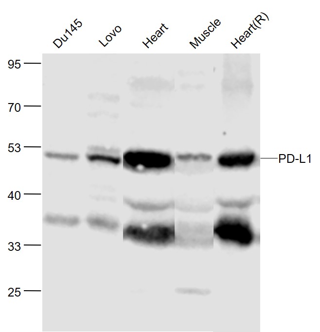 Sample:
Du145(Human) Cell Lysate at 30 ug
Lovo(Human) Cell Lysate at 30 ug
Heart(Mouse) Lysate at 40 ug
Muscle(Mouse) Lysate at 40 ug
Heart(Rat) Lysate at 40 ug
Primary: Anti-PD-L1 (SL10159R) at 1/500 dilution
Secondary: IRDye800CW Goat Anti-Rabbit IgG at 1/20000 dilution
Predicted band size: 50 kD
Observed band size: 50 kD
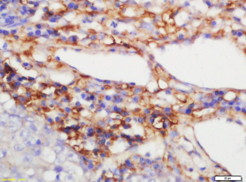 Tissue/cell: human lung carcinoma; 4% Paraformaldehyde-fixed and paraffin-embedded;
Antigen retrieval: citrate buffer ( 0.01M, pH 6.0 ), Boiling bathing for 15min; Block endogenous peroxidase by 3% Hydrogen peroxide for 30min; Blocking buffer (normal goat serum,C-0005) at 37℃ for 20 min;
Incubation: Anti-CD274 Polyclonal Antibody, Unconjugated(SL10159R) 1:200, overnight at 4°C, followed by conjugation to the secondary antibody(SP-0023) and DAB(C-0010) staining
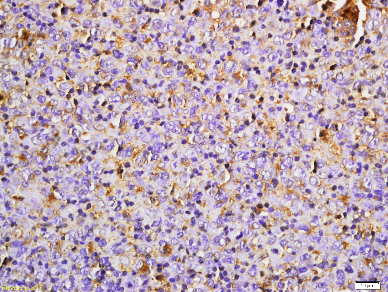 Tissue/cell: mouse transplantable tumor; 4% Paraformaldehyde-fixed and paraffin-embedded;
Antigen retrieval: citrate buffer ( 0.01M, pH 6.0 ), Boiling bathing for 15min; Block endogenous peroxidase by 3% Hydrogen peroxide for 30min; Blocking buffer (normal goat serum,C-0005) at 37℃ for 20 min;
Incubation: Anti-CD274 Polyclonal Antibody, Unconjugated(SL10159R) 1:100, overnight at 4°C, followed by conjugation to the secondary antibody(SP-0023) and DAB(C-0010) staining
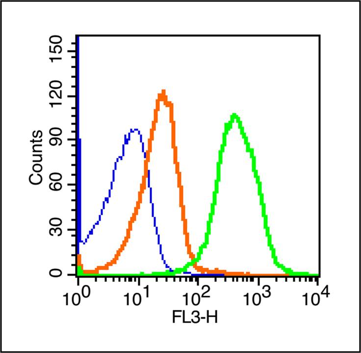 Blank control (blue line): Mouse spleen cells(fixed with 70% ice-cold methanol overnight at 4℃).
Primary Antibody (green line): Rabbit Anti-CD274/PE-CY7 Conjugated antibody (SL10159R-PE-CY7),Dilution: 1μg /10^6 cells;
Isotype Control Antibody (orange line): Rabbit IgG-PE-CY7 .
 Blank control: Hela.
Primary Antibody (green line): Rabbit Anti-PD-L1 antibody (SL10159R)
Dilution: 2ug/Test;
Secondary Antibody : Goat anti-rabbit IgG-FITC
Dilution: 0.5ug/Test.
Protocol
The cells were incubated in 5%BSA to block non-specific protein-protein interactions for 30 min at room temperature .Cells stained with Primary Antibody for 30 min at room temperature. The secondary antibody used for 40 min at room temperature. Acquisition of 20,000 events was performed.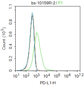 Blank control: Hela.
Primary Antibody (green line): Rabbit Anti-PD-L1 antibody (SL10159R)
Dilution: 2ug/Test;
Secondary Antibody : Goat anti-rabbit IgG-FITC
Dilution: 0.5ug/Test.
Protocol
The cells were incubated in 5%BSA to block non-specific protein-protein interactions for 30 min at room temperature .Cells stained with Primary Antibody for 30 min at room temperature. The secondary antibody used for 40 min at room temperature. Acquisition of 20,000 events was performed.
Cartpieces
Totalgoods,subtotals:¥Checkout
Partial purchase records(bought amounts latest0)
No one bought this product
User Comment(Total0User Comment Num)
- No comment


 +86 571 56623320


