
Rabbit Anti-Runx3 antibody
RUNX3_HUMAN; Runt-related transcription factor 3; AML2; CBFA3; PEBP2A3; Acute myeloid leukemia 2 protein; Core-binding factor subunit alpha-3 (CBF-alpha-3); Oncogene AML-2; Polyomavirus enhancer-binding protein 2 alpha C subunit (PEA2-alpha C; PEBP2-alpha
View History [Clear]
Details
Product Name Runx3 Chinese Name Runx3抗体 Alias RUNX3_HUMAN; Runt-related transcription factor 3; AML2; CBFA3; PEBP2A3; Acute myeloid leukemia 2 protein; Core-binding factor subunit alpha-3 (CBF-alpha-3); Oncogene AML-2; Polyomavirus enhancer-binding protein 2 alpha C subunit (PEA2-alpha C; PEBP2-alpha C); SL3-3 enhancer factor 1 alpha C subunit; SL3/AKV core-binding factor alpha C subunit; literatures Research Area Tumour Cell biology Immunogen Species Rabbit Clonality Polyclonal React Species Human, Mouse, (predicted: Rat, Chicken, Dog, Cow, ) Applications WB=1:500-2000 ELISA=1:5000-10000 IHC-P=1:100-500 IHC-F=1:100-500 IF=1:100-500 (Paraffin sections need antigen repair)
not yet tested in other applications.
optimal dilutions/concentrations should be determined by the end user.Theoretical molecular weight 44kDa Cellular localization The nucleus cytoplasmic Form Liquid Concentration 1mg/ml immunogen KLH conjugated synthetic peptide derived from human Runx3: 151-250/415 Lsotype IgG Purification affinity purified by Protein A Buffer Solution 0.01M TBS(pH7.4) with 1% BSA, 0.03% Proclin300 and 50% Glycerol. Storage Shipped at 4℃. Store at -20 °C for one year. Avoid repeated freeze/thaw cycles. Attention This product as supplied is intended for research use only, not for use in human, therapeutic or diagnostic applications. PubMed PubMed Product Detail This gene encodes a member of the runt domain-containing family of transcription factors. A heterodimer of this protein and a beta subunit forms a complex that binds to the core DNA sequence 5'-PYGPYGGT-3' found in a number of enhancers and promoters, and can either activate or suppress transcription. It also interacts with other transcription factors. It functions as a tumor suppressor, and the gene is frequently deleted or transcriptionally silenced in cancer. Multiple transcript variants encoding different isoforms have been found for this gene. [provided by RefSeq, Jul 2008]
Function:
CBF binds to the core site, 5'-PYGPYGGT-3', of a number of enhancers and promoters, including murine leukemia virus, polyomavirus enhancer, T-cell receptor enhancers, lck, IL-3 and GM-CSF promoters.
Subunit:
Heterodimer of an alpha and a beta subunit. The alpha subunit binds DNA as a monomer and through the Runt domain. DNA-binding is increased by heterodimerization. Interacts with TLE1 and SUV39H1. The tyrosine phosphorylated form (via runt domain) interacts with SRC (via protein kinase domain). Interacts with FYN and LCK.
Subcellular Location:
Nucleus. Cytoplasm. Note=The tyrosine phosphorylated form localizes to the cytoplasm.
Post-translational modifications:
Phosphorylated on tyrosine residues by SRC. Phosphorylated by LCK and FYN.
Similarity:
Contains 1 Runt domain.
SWISS:
Q13761
Gene ID:
864
Database links:Entrez Gene: 864 Human
Omim: 600210 Human
SwissProt: Q13761 Human
Unigene: 170019 Human
新型抑癌基因/一种新发现的抑癌基因。经研究发现,在人类胃癌细胞系和胃癌组织中普遍存在Runx3基因表达的缺失或下调,该蛋白与胃癌的发生、发展密切相关。
RUNX3基因被认为是一种新发现的抑癌基因,对胃癌细胞的生长有明显的抑制作用在胃黏膜epithelial cells调控、对脊神经节的神经发育和TCell differentiation中都发挥重要作用。
随着研究的不断深入,Runx3蛋白有望成为胃癌诊断的一个新型生物学Maker和基因治疗靶点。Product Picture 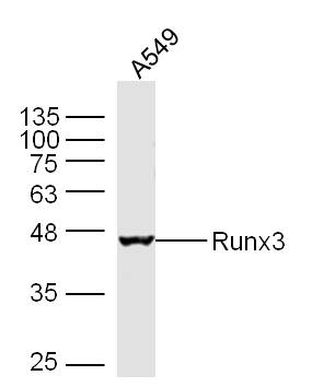 Sample: A549 Cell Lysate at 30 ug
Primary: Anti- Runx3 (SL0378R) at 1/300 dilution
Secondary: IRDye800CW Goat Anti-Mouse IgG at 1/20000 dilution
Predicted band size: 44 kD
Observed band size: 44 kD
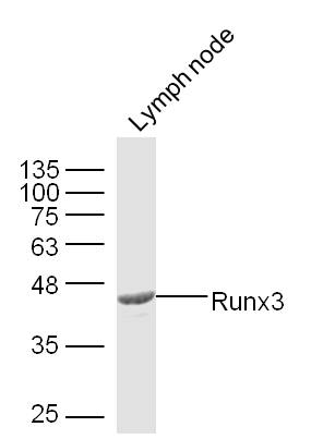 Sample: Lymph node (Mouse) Lysate at 30 ug
Primary: Anti- Runx3 (SL0378R) at 1/300 dilution
Secondary: IRDye800CW Goat Anti-Mouse IgG at 1/20000 dilution
Predicted band size: 44 kD
Observed band size: 44 kD
 Tissue/cell: human cervical carcinoma; 4% Paraformaldehyde-fixed and paraffin-embedded;
Antigen retrieval: citrate buffer ( 0.01M, pH 6.0 ), Boiling bathing for 15min; Block endogenous peroxidase by 3% Hydrogen peroxide for 30min; Blocking buffer (normal goat serum,C-0005) at 37℃ for 20 min;
Incubation: Anti-Runx3 Polyclonal Antibody, Unconjugated(SL0378R) 1:300, overnight at 4°C, followed by conjugation to the secondary antibody(SP-0023) and DAB(C-0010) staining
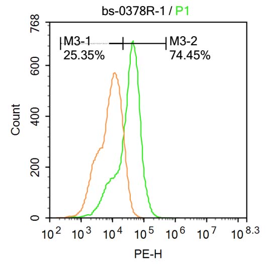 U-937 cells were fixed with 4% PFA for 10min at room temperature,permeabilized with 90% ice-cold methanol for 20 min at room temperature,and incubated in 5% BSA blocking buffer for 30 min at room temperature. Cells were then stained with Runx3 Antibody(SL0378R) at 1:500 dilution in blocking buffer and incubated for 30 min at room temperature, washed twice with 2%BSA in PBS, followed by secondary antibody incubation for 40 min at room temperature. Acquisitions of 20,000 events were performed.Cells stained with primary antibody (green), and isotype control (orange).
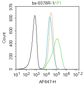 Blank control: Jurkat.
Primary Antibody (green line): Rabbit Anti-Runx3 antibody (SL0378R)
Dilution: 1μg /10^6 cells;
Isotype Control Antibody (orange line): Rabbit IgG .
Secondary Antibody : Goat anti-rabbit IgG-AF647
Dilution: 1μg /test.
Protocol
The cells were fixed with 4% PFA (10min at room temperature)and then permeabilized with 90% ice-cold methanol for 20 min at-20℃. The cells were then incubated in 5%BSA to block non-specific protein-protein interactions for 30 min at room temperature .Cells stained with Primary Antibody for 30 min at room temperature. The secondary antibody used for 40 min at room temperature. Acquisition of 20,000 events was performed.
Cartpieces
Totalgoods,subtotals:¥Checkout
Partial purchase records(bought amounts latest0)
No one bought this product
User Comment(Total0User Comment Num)
- No comment


 +86 571 56623320


