
Rabbit Anti-PDE7B antibody
PDE 7B; PDE7B protein; Phosphodiesterase 7B; Phosphodiesterase7B; RNPDE7B; Rolipram insensitive phosphodiesterase type 7; bA472E5.1; cAMP specific 3 5 cyclic phosphodiesterase 7B; cAMP specific 3'5' cyclic phosphodiesterase 7B; High affinity cAMP specific
View History [Clear]
Details
Product Name PDE7B Chinese Name 磷酸二酯酶7B抗体 Alias PDE 7B; PDE7B protein; Phosphodiesterase 7B; Phosphodiesterase7B; RNPDE7B; Rolipram insensitive phosphodiesterase type 7; bA472E5.1; cAMP specific 3 5 cyclic phosphodiesterase 7B; cAMP specific 3'5' cyclic phosphodiesterase 7B; High affinity cAMP specific 3 5 cyclic phosphodiesterase; High affinity cAMP specific 3'5' cyclic phosphodiesterase; PDE7B_HUMAN. Research Area Neurobiology Signal transduction The cell membrane受体 G protein-coupled receptor G protein signal Immunogen Species Rabbit Clonality Polyclonal React Species Human, Mouse, Rat, (predicted: Dog, Pig, Cow, Horse, Sheep, ) Applications WB=1:500-2000 ELISA=1:5000-10000 IHC-P=1:100-500 IHC-F=1:100-500 Flow-Cyt=1μg/Test ICC=1:100-500 IF=1:100-500 (Paraffin sections need antigen repair)
not yet tested in other applications.
optimal dilutions/concentrations should be determined by the end user.Theoretical molecular weight 52kDa Cellular localization cytoplasmic Form Liquid Concentration 1mg/ml immunogen KLH conjugated synthetic peptide derived from human PDE7B: 281-360/450 Lsotype IgG Purification affinity purified by Protein A Buffer Solution 0.01M TBS(pH7.4) with 1% BSA, 0.03% Proclin300 and 50% Glycerol. Storage Shipped at 4℃. Store at -20 °C for one year. Avoid repeated freeze/thaw cycles. Attention This product as supplied is intended for research use only, not for use in human, therapeutic or diagnostic applications. PubMed PubMed Product Detail Phosphodiesterases (PDE, also designated cyclic nucleotide phosphodiesterase) are important for the downregulation of the intracellular level of the second messenger cyclic adenosine monophosphate (cAMP) by hydrolyzing cAMP to 5'AMP. Phosphodiesterase type 3 isoforms, PDE3A and 3B, are expressed primarily in cardiovascular tissue and adipose tissue, respectively. PDE3A, is found in myocardium and platelets and PDE3B is found in lymphocytes. The PDE7A1 (HCP1) isozyme and the PDE7A2 proteins, alternate splice products of PDE7A, are highly expressed in skeletal muscle. PDE7B is most highly expressed in pancreas. The PDE family contains proteins that serve tissue-specific roles in regulation of lipolysis, glycogenolysis, myocardial contractility, and smooth muscle relaxation.
Function:
PDE7B is a cAMP specific phosphodiesterase. The cAMP-specific phosphodiesterase type-7 (PDE7) family is comprised of 2 genes (PDE7A and PDE7B) each with multiple splice variants generated by RNA splicing and use of alternate initiation sites. PDE7B has three splice variants (PDE7B1 = 446 amino acid; PDE7B2 =359 amino acids and PDE7B3 = 459 amino acids. Both PDE7B2 ad PDE7B3 posses unique N-terminal sequences. PDE7B is expressed in various tissues including skeletal muscle, heart, lung and testis. PDE7B may be involved in the control of cAMP-mediated neural activity and cAMP metabolism in the brain.
Tissue Specificity:
Highly expressed in brain. Also expressed in heart, liver, skeletal muscle and pancreas.
Similarity:
Belongs to the cyclic nucleotide phosphodiesterase family. PDE7 subfamily.
SWISS:
Q9NP56
Gene ID:
27115
Database links:Entrez Gene: 27115 Human
Entrez Gene: 29863 Mouse
Omim: 604645 Human
SwissProt: Q9NP56 Human
SwissProt: Q9QXQ1 Mouse
Unigene: 153615 Rat
Product Picture 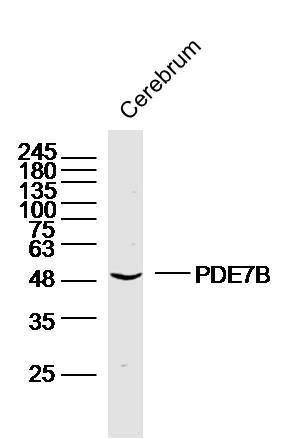 Sample: Cerebrum (Mouse) Lysate at 40 ug
Primary: Anti-PDE7B (SL11576R) at 1/300 dilution
Secondary: IRDye800CW Goat Anti-Rabbit IgG at 1/20000 dilution
Predicted band size: 52 kD
Observed band size: 52 kD
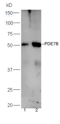 Sample:
Brain (Mouse) Lysate at 40 ug
Liver (Mouse) Lysate at 40 ug
Primary: Anti-PDE7B (SL11576R) at 1/300 dilution
Secondary: HRP conjugated Goat-Anti-rabbit IgG (SL0295G-HRP) at 1/5000 dilution
Predicted band size: 52 kD
Observed band size: 52 kD
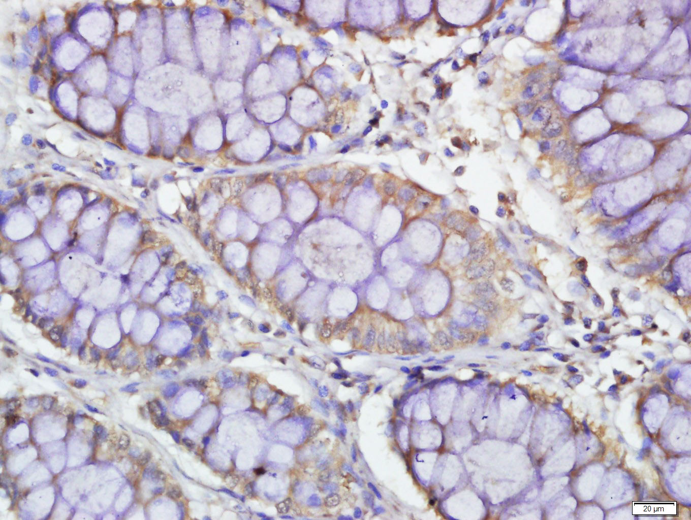 Paraformaldehyde-fixed, paraffin embedded (human colon carcinoma); Antigen retrieval by boiling in sodium citrate buffer (pH6.0) for 15min; Block endogenous peroxidase by 3% hydrogen peroxide for 20 minutes; Blocking buffer (normal goat serum) at 37∑C for 30min; Antibody incubation with (PDE7B) Polyclonal Antibody, Unconjugated (SL11576R) at 1:500 overnight at 4∑C, followed by a conjugated secondary (sp-0023) for 20 minutes and DAB staining.
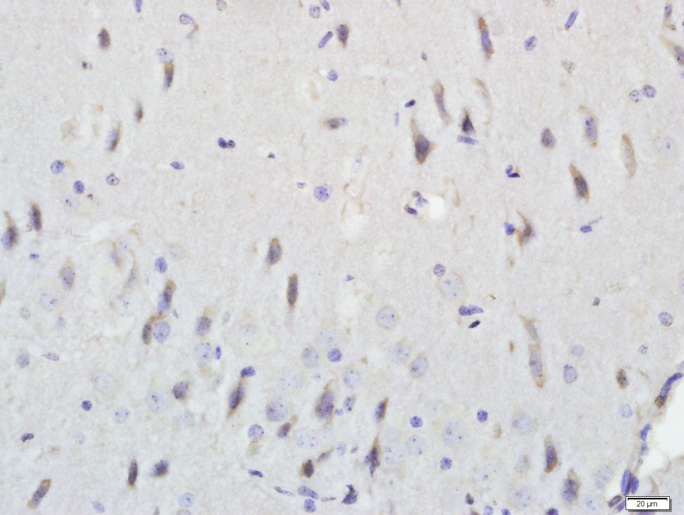 raformaldehyde-fixed, paraffin embedded (rat brain); Antigen retrieval by boiling in sodium citrate buffer (pH6.0) for 15min; Block endogenous peroxidase by 3% hydrogen peroxide for 20 minutes; Blocking buffer (normal goat serum) at 37∑C for 30min; Antibody incubation with (PDE7B) Polyclonal Antibody, Unconjugated (SL11576R) at 1:500 overnight at 4∑C, followed by a conjugated secondary (sp-0023) for 20 minutes and DAB staining.
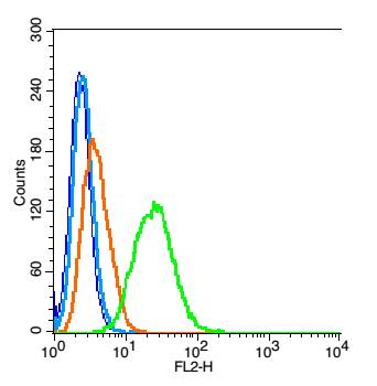 Blank control(blue): RSC96 cells(fixed with 2% paraformaldehyde (10 min) , then permeabilized with 90% ice-cold methanol for 30 min on ice).
Primary Antibody:Rabbit Anti- PDE7B antibody(SL11576R), Dilution: 1ug in 100 uL 1X PBS containing 0.5% BSA;
Isotype Control Antibody: Rabbit IgG(orange) ,used under the same conditions );
Secondary Antibody: Goat anti-rabbit IgG-PE(white blue), Dilution: 1:200 in 1 X PBS containing 0.5% BSA.
Cartpieces
Totalgoods,subtotals:¥Checkout
Bought notes(bought amounts latest0)
No one bought this product
User Comment(Total0User Comment Num)
- No comment


 +86 571 56623320
 +86 18668110335

