
Rabbit Anti-VAX1 antibody
VAX1; VAX1_HUMAN; ventral anterior homeobox 1.
View History [Clear]
Details
Product Name VAX1 Chinese Name 视神经视网膜相关蛋白VAX1抗体 Alias VAX1; VAX1_HUMAN; ventral anterior homeobox 1. Research Area Cell biology Neurobiology Epigenetics Immunogen Species Rabbit Clonality Polyclonal React Species Human, Mouse, Rat, (predicted: Chicken, Dog, Pig, Cow, ) Applications WB=1:500-2000 ELISA=1:5000-10000 Flow-Cyt=1μg/Test ICC=1:100
not yet tested in other applications.
optimal dilutions/concentrations should be determined by the end user.Theoretical molecular weight 35kDa Cellular localization The nucleus Form Liquid Concentration 1mg/ml immunogen KLH conjugated synthetic peptide derived from human VAX1: 133-200/334 Lsotype IgG Purification affinity purified by Protein A Buffer Solution 0.01M TBS(pH7.4) with 1% BSA, 0.03% Proclin300 and 50% Glycerol. Storage Shipped at 4℃. Store at -20 °C for one year. Avoid repeated freeze/thaw cycles. Attention This product as supplied is intended for research use only, not for use in human, therapeutic or diagnostic applications. PubMed PubMed Product Detail The homeobox DNA-binding domain is a 60 amino acid motif that is conserved among many species and functions to bind DNA via a helix-turn-helix structure, thereby playing a role in transcriptional regulation and the control of gene expression. VAX1 (ventral anterior homeobox 1) is a 334 amino acid protein that localizes to the nucleus and contains one homeobox DNA-binding domain. Expressed as multiple alternatively spliced isoforms, VAX1 is required for major tract formation and axon guidance in the developing brain and may play a role in the differentiation of various structures, including the optic stalk, the neuroretina and the pigmented epithelium. The gene encoding VAX1 maps to human chromosome 10, which houses over 1,200 genes and comprises nearly 4.5% of the human genome.
Function:
Required for axon guidance and major tract formation in the developing forebrain. May contribute to the differentiation of the neuroretina, pigmented epithelium and optic stalk.
Subcellular Location:
Nucleus.
DISEASE:
Defects in VAX1 are the cause of microphthalmia, syndromic, type 11 (MCOPS11) [MIM:614402]. A rare clinical entity including as main characteristics microphthalmia and small optic nerves, cleft lip and palate, absence of corpus callosum, hippocampal malformations, and absence of the pineal gland. Microphthalmia is a disorder of eye formation, ranging from small size of a single eye to complete bilateral absence of ocular tissues (anophthalmia). In many cases, microphthalmia/anophthalmia occurs in association with syndromes that include non-ocular abnormalities.
Similarity:
Belongs to the EMX homeobox family.
Contains 1 homeobox DNA-binding domain.
SWISS:
Q5SQQ9
Gene ID:
11023
Database links:Entrez Gene: 11023 Human
Entrez Gene: 22326 Mouse
Omim: 604294 Human
SwissProt: Q5SQQ9 Human
SwissProt: Q2NKI2 Mouse
Unigene: 441536 Human
Unigene: 23801 Mouse
Unigene: 48764 Rat
Product Picture 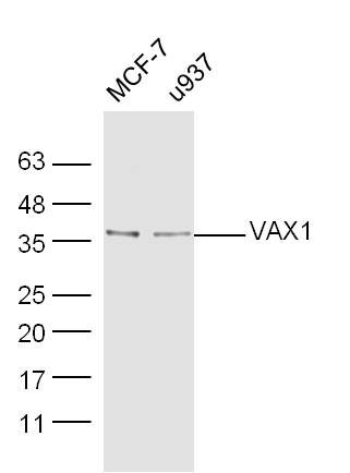 Sample:
MCF-7 Cell (Human) Lysate at 40 ug
U937 Cell (Human) Lysate at 40 ug
Primary: Anti-VAX1 (SL11496R) at 1/300 dilution
Secondary: IRDye800CW Goat Anti-Rabbit IgG at 1/20000 dilution
Predicted band size: 35 kD
Observed band size: 36 kD
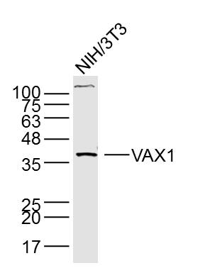 Sample: NIH/3T3 Cell (Mouse) Lysate at 40 ug
Primary: Anti- VAX1 (SL11496R) at 1/300 dilution
Secondary: IRDye800CW Goat Anti-Rabbit IgG at 1/20000 dilution
Predicted band size: 35 kD
Observed band size: 37 kD
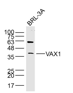 Sample: BRL-3A Cell (Rat) Lysate at 40 ug
Primary: Anti- VAX1 (SL11496R) at 1/300 dilution
Secondary: IRDye800CW Goat Anti-Rabbit IgG at 1/20000 dilution
Predicted band size: 35 kD
Observed band size: 37 kD
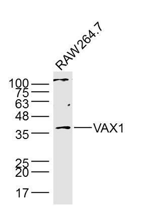 Sample: RAW264.7 Cell (Mouse) Lysate at 40 ug
Primary: Anti- VAX1 (SL11496R) at 1/300 dilution
Secondary: IRDye800CW Goat Anti-Rabbit IgG at 1/20000 dilution
Predicted band size: 35 kD
Observed band size: 37 kD
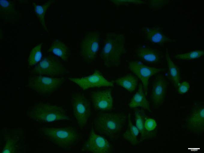 A549 cell; 4% Paraformaldehyde-fixed; Triton X-100 at room temperature for 20 min; Blocking buffer (normal goat serum, C-0005) at 37°C for 20 min; Antibody incubation with (VAX1) polyclonal Antibody, Unconjugated (SL11496R) 1:100, 90 minutes at 37°C; followed by a conjugated Goat Anti-Rabbit IgG antibody at 37°C for 90 minutes, DAPI (blue, C02-04002) was used to stain the cell nuclei.
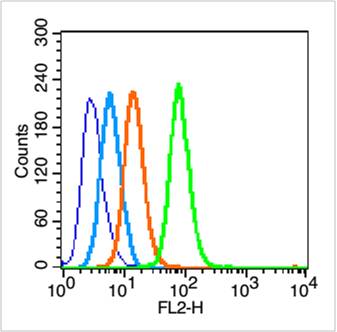 Blank control (blue line): Hep G2 (fixed with 70% ethanol (Overnight at 4℃) and then permeabilized with 90% methanol for 20 min at -20℃).
Primary Antibody (green line): Rabbit Anti-VAX1 antibody (SL11496R),Dilution: 0.2μg /10^6 cells;
Isotype Control Antibody (orange line): Rabbit IgG .
Secondary Antibody (white blue line): Goat anti-rabbit IgG-PE,Dilution: 1μg /test.
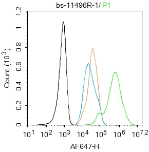 Blank control:A431.
Primary Antibody (green line): Rabbit Anti-VAX1 antibody (SL11496R)
Dilution: 1μg /10^6 cells;
Isotype Control Antibody (orange line): Rabbit IgG .
Secondary Antibody : Goat anti-rabbit IgG-AF647
Dilution: 1μg /test.
Protocol
The cells were fixed with 4% PFA (10min at room temperature)and then permeabilized with 90% ice-cold methanol for 20 min at -20℃.The cells were then incubated in 5%BSA to block non-specific protein-protein interactions for 30 min at room temperature.Cells stained with Primary Antibody for 30 min at room temperature. The secondary antibody used for 40 min at room temperature. Acquisition of 20,000 events was performed.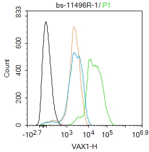 Blank control(black line):A549.
Primary Antibody (green line): Rabbit Anti-VAX1 antibody (SL11496R)
Dilution:1ug/Test;
Secondary Antibody(white blue line): Goat anti-rabbit IgG-AF488
Dilution: 0.5ug/Test.
Isotype control(orange line): Normal Rabbit IgG
Protocol
The cells were fixed with 4% PFA (10min at room temperature)and then permeabilized with 90% ice-cold methanol for 20 min at -20℃, The cells were then incubated in 5%BSA to block non-specific protein-protein interactions for 30 min at room temperature .Cells stained with Primary Antibody for 30 min at room temperature. The secondary antibody used for 40 min at room temperature. Acquisition of 20,000 events was performed.
Cartpieces
Totalgoods,subtotals:¥Checkout
Bought notes(bought amounts latest0)
No one bought this product
User Comment(Total0User Comment Num)
- No comment


 +86 571 56623320
 +86 18668110335

