
Rabbit Anti-THRB1 antibody
Thyroid Hormone Receptor beta; Avian erythroblastic leukemia viral (v erb a) oncogene homolog 2; C ERBA 2; C ERBA BETA; c-erbA-2; c-erbA-beta; ERBA 2; ERBA BETA; ERBA2; Erythroblastic leukemia viral (v erb a) oncogene homolog 2 avian; generalized resistan
View History [Clear]
Details
Product Name THRB1 Chinese Name 甲状腺激素受体β抗体 Alias Thyroid Hormone Receptor beta; Avian erythroblastic leukemia viral (v erb a) oncogene homolog 2; C ERBA 2; C ERBA BETA; c-erbA-2; c-erbA-beta; ERBA 2; ERBA BETA; ERBA2; Erythroblastic leukemia viral (v erb a) oncogene homolog 2 avian; generalized resistance to thyroid hormone; GRTH; MGC126109; MGC126110; NR1A2; Nuclear receptor subfamily 1 group A member 2; Oncogene ERBA2; PRTH; THB_HUMAN; THR 1; THR1; THRB 1; THRB 2; THRB; THRB2; Thyroid hormone nuclear receptor beta variant 1; Thyroid hormone receptor beta 1; Thyroid hormone receptor beta 2; Thyroid hormone receptor beta; Thyroid hormone receptor, beta (erythroblastic leukemia viral (v erb a) oncogene homolog 2, avian); THβ1; THβ2. Research Area Cardiovascular Cell biology Neurobiology Growth factors and hormones Endocrinopathy Immunogen Species Rabbit Clonality Polyclonal React Species Human, Mouse, Rat, (predicted: Chicken, Cow, Rabbit, Sheep, ) Applications WB=1:500-2000 ELISA=1:5000-10000 IHC-P=1:100-500 IHC-F=1:100-500 Flow-Cyt=1ug/Test IF=1:100-500 (Paraffin sections need antigen repair)
not yet tested in other applications.
optimal dilutions/concentrations should be determined by the end user.Theoretical molecular weight 53 kDa Cellular localization The nucleus Form Liquid Concentration 1mg/ml immunogen KLH conjugated synthetic peptide derived from human Thyroid Hormone Receptor beta: 201-300/461 Lsotype IgG Purification affinity purified by Protein A Buffer Solution 0.01M TBS(pH7.4) with 1% BSA, 0.03% Proclin300 and 50% Glycerol. Storage Shipped at 4℃. Store at -20 °C for one year. Avoid repeated freeze/thaw cycles. Attention This product as supplied is intended for research use only, not for use in human, therapeutic or diagnostic applications. PubMed PubMed Product Detail Thyroid hormone receptors (TRs) are ligand-dependent transcription factors that mediate the biological activities of thyroid hormone (T3). Thyroid hormone receptor b2 (TRb2) is a high affinity receptor for triiodothyronine which belongs to the nuclear hormone receptor family and the NR1 subfamily. It is composed of three domains: a modulating N-terminal domain, a DNA-binding domain and a C-terminal steroid-binding domain. Defects in the receptor result in generalized thyroid hormone resistance (GTHR). GTHR is transmitted as an autosomal dominant trait, but an autosomal recessive form also exists. The disease is characterized by goiter, abnormal mental functions, increased susceptibility to infections, abnormal growth and bone maturation, tachycardia and deafness. GTHR patients also have high levels of circulating thyroid hormones (T3-T4), with normal or slightly elevated thyroid stimulating hormone.
Function:
High affinity receptor for triiodothyronine.
Subunit:
Binds DNA as a dimer; homodimer and heterodimer with RXRB. Interacts with NCOA7 in a ligand-inducible manner. Interacts with C1D. Interacts with NR2F6; the interaction impairs the binding of the THRB homodimer and THRB:RXRB heterodimer to T3 response elements. Interacts with PRMT2 and THRSP.
Subcellular Location:
Nucleus.
DISEASE:
Defects in THRB are the cause of generalized thyroid hormone resistance (GTHR) [MIM:188570, 274300]. GTHR is transmitted as an autosomal dominant trait, but an autosomal recessive form also exists. The disease is characterized by goiter, abnormal mental functions, increased susceptibility to infections, abnormal growth and bone maturation, tachycardia and deafness. Affected individuals may also have attention deficit-hyperactivity disorders (ADHD) and language difficulties. GTHR patients also have high levels of circulating thyroid hormones (T3-T4), with normal or slightly elevated thyroid stimulating hormone (TSH).
Similarity:
Belongs to the nuclear hormone receptor family. NR1 subfamily.
Contains 1 nuclear receptor DNA-binding domain.
SWISS:
P10828
Gene ID:
7068
Database links:Entrez Gene: 7068 Human
Omim: 190160 Human
SwissProt: P10828 Human
Unigene: 187861 Human
Unigene: 728126 Human
Unigene: 88692 Rat
Product Picture 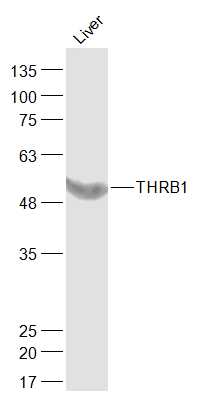 Sample:
Liver (Mouse) Lysate at 40 ug
Primary: Anti-THRB1 (SL11440R) at 1/1000 dilution
Secondary: IRDye800CW Goat Anti-Rabbit IgG at 1/20000 dilution
Predicted band size: 53 kD
Observed band size: 53 kD
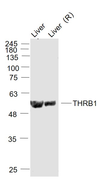 Sample:
Liver (Mouse) Lysate at 40 ug
Liver (Rat) Lysate at 40 ug
Primary: Anti- THRB1 (SL11440R) at 1/2000 dilution
Secondary: IRDye800CW Goat Anti-Rabbit IgG at 1/20000 dilution
Predicted band size: 53 kD
Observed band size: 53 kD
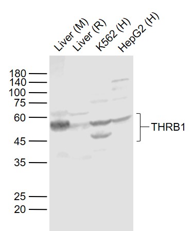 Sample:
Lane 1: Liver (Mouse) Lysate at 40 ug
Lane 2: Liver (Rat) Lysate at 40 ug
Lane 3: K562 (Human) Cell Lysate at 30 ug
Lane 4: HepG2 (Human) Cell Lysate at 30 ug
Primary: Anti- THRB1 (SL11440R) at 1/1000 dilution
Secondary: IRDye800CW Goat Anti-Rabbit IgG at 1/20000 dilution
Predicted band size: 53/46 kD
Observed band size: 53/46 kD
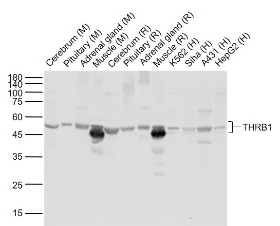 Sample:
Lane 1: Cerebrum (Mouse) Lysate at 40 ug
Lane 2: Pituitary (Mouse) Lysate at 40 ug
Lane 3: Adrenal gland (Mouse) Lysate at 40 ug
Lane 4: Muscle (Mouse) Lysate at 40 ug
Lane 5: Cerebrum (Rat) Lysate at 40 ug
Lane 6: Pituitary (Rat) Lysate at 40 ug
Lane 7: Adrenal gland (Rat) Lysate at 40 ug
Lane 8: Muscle (Rat) Lysate at 40 ug
Lane 9: K562 (Human) Cell Lysate at 30 ug
Lane 10: Siha (Human) Cell Lysate at 30 ug
Lane 11: A431 (Human) Cell Lysate at 30 ug
Lane 12: HepG2 (Human) Cell Lysate at 30 ug
Primary: Anti-THRB1 (SL11440R) at 1/1000 dilution
Secondary: IRDye800CW Goat Anti-Rabbit IgG at 1/20000 dilution
Predicted band size: 53/46 kD
Observed band size: 53/46 kD
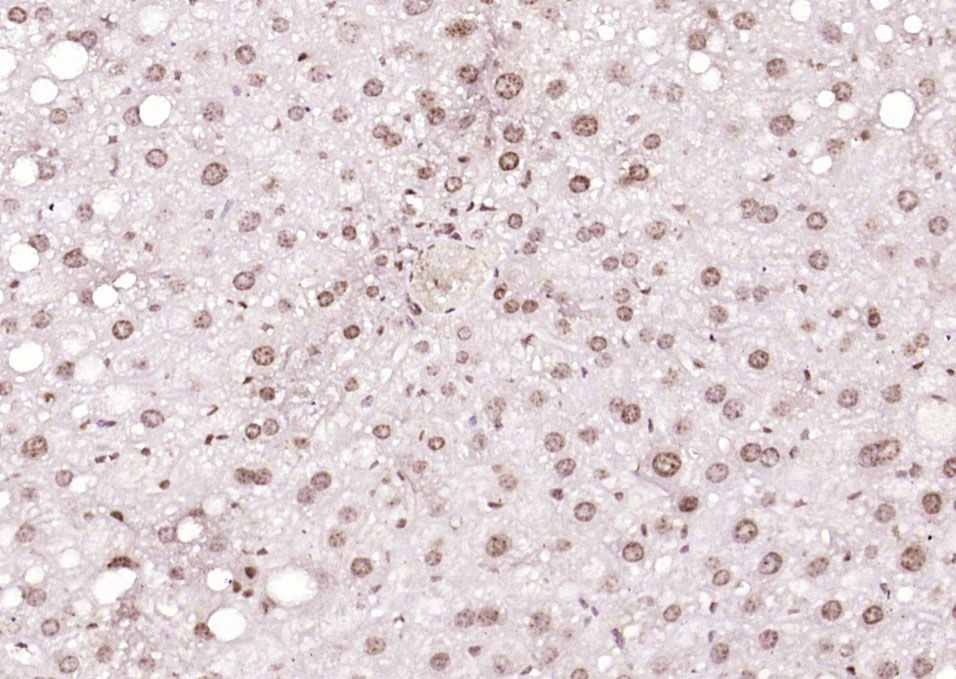 Paraformaldehyde-fixed, paraffin embedded (mouse liver); Antigen retrieval by boiling in sodium citrate buffer (pH6.0) for 15min; Block endogenous peroxidase by 3% hydrogen peroxide for 20 minutes; Blocking buffer (normal goat serum) at 37°C for 30min; Antibody incubation with (THRB1) Polyclonal Antibody, Unconjugated (SL11440R) at 1:200 overnight at 4°C, followed by operating according to SP Kit(Rabbit) (sp-0023) instructionsand DAB staining.
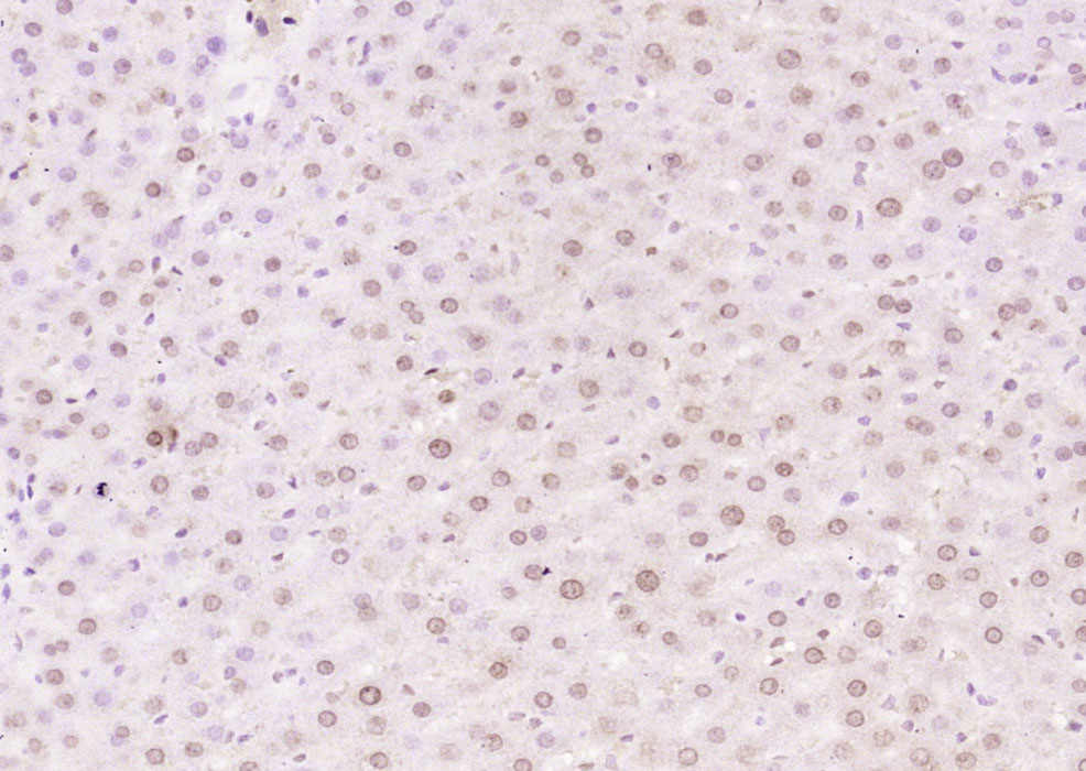 Paraformaldehyde-fixed, paraffin embedded (rat liver); Antigen retrieval by boiling in sodium citrate buffer (pH6.0) for 15min; Block endogenous peroxidase by 3% hydrogen peroxide for 20 minutes; Blocking buffer (normal goat serum) at 37°C for 30min; Antibody incubation with (THRB1) Polyclonal Antibody, Unconjugated (SL11440R) at 1:200 overnight at 4°C, followed by operating according to SP Kit(Rabbit) (sp-0023) instructionsand DAB staining.
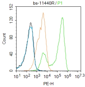 Blank control: HepG2.
Primary Antibody (green line): Rabbit Anti-THRB1 antibody (SL11440R)
Dilution: 1μg /10^6 cells;
Isotype Control Antibody (orange line): Rabbit IgG .
Secondary Antibody : Goat anti-rabbit IgG-PE
Dilution: 1μg /test.
Protocol
The cells were fixed with 4% PFA (10min at room temperature)and then permeabilized with 90% ice-cold methanol for 20 min at-20℃. The cells were then incubated in 5%BSA to block non-specific protein-protein interactions for 30 min at at room temperature .Cells stained with Primary Antibody for 30 min at room temperature. The secondary antibody used for 40 min at room temperature. Acquisition of 20,000 events was performed.Blank control: HepG2.
Primary Antibody (green line): Rabbit Anti-THRB1 antibody (SL11440R)
Dilution: 1μg /10^6 cells;
Isotype Control Antibody (orange line): Rabbit IgG .
Secondary Antibody : Goat anti-rabbit IgG-PE
Dilution: 1μg /test.
Protocol
The cells were fixed with 4% PFA (10min at room temperature)and then permeabilized with 90% ice-cold methanol for 20 min at-20℃. The cells were then incubated in 5%BSA to block non-specific protein-protein interactions for 30 min at at room temperature .Cells stained with Primary Antibody for 30 min at room temperature. The secondary antibody used for 40 min at room temperature. Acquisition of 20,000 events was performed.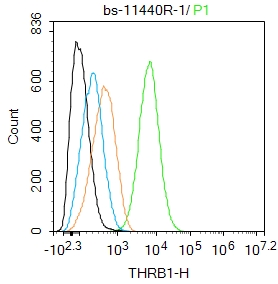 Blank control: A431.
Primary Antibody (green line): Rabbit Anti-THRB1 antibody (SL 11440R)
Dilution: 1ug/Test;
Secondary Antibody : Goat anti-rabbit IgG-FITC
Dilution: 0.5ug/Test.
Protocol
The cells were fixed with 4% PFA (10min at room temperature)and then permeabilized with 90% ice-cold methanol for 20 min at -20℃.The cells were then incubated in 5%BSA to block non-specific protein-protein interactions for 30 min at room temperature .Cells stained with Primary Antibody for 30 min at room temperature. The secondary antibody used for 40 min at room temperature. Acquisition of 20,000 events was performed.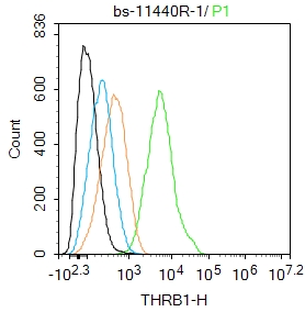 Blank control: A431.
Primary Antibody (green line): Rabbit Anti-THRB1 antibody (SL 11440R)
Dilution: 1ug/Test;
Secondary Antibody : Goat anti-rabbit IgG-FITC
Dilution: 0.5ug/Test.
Protocol
The cells were fixed with 4% PFA (10min at room temperature)and then permeabilized with 90% ice-cold methanol for 20 min at -20℃.The cells were then incubated in 5%BSA to block non-specific protein-protein interactions for 30 min at room temperature .Cells stained with Primary Antibody for 30 min at room temperature. The secondary antibody used for 40 min at room temperature. Acquisition of 20,000 events was performed.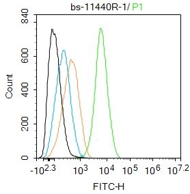 Blank control: A431.
Primary Antibody (green line): Rabbit Anti-THRB1 antibody (SL 11440R)
Dilution: 1ug/Test;
Secondary Antibody : Goat anti-rabbit IgG-FITC
Dilution: 0.5ug/Test.
Protocol
The cells were fixed with 4% PFA (10min at room temperature)and then permeabilized with 90% ice-cold methanol for 20 min at -20℃.The cells were then incubated in 5%BSA to block non-specific protein-protein interactions for 30 min at room temperature .Cells stained with Primary Antibody for 30 min at room temperature. The secondary antibody used for 40 min at room temperature. Acquisition of 20,000 events was performed.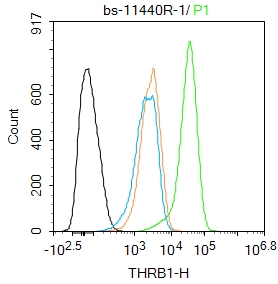 Blank control:A431.
Primary Antibody (green line): Rabbit Anti-THRB1 antibody (SL11440R)
Dilution: 1ug/Test;
Secondary Antibody : Goat anti-rabbit IgG-AF488
Dilution: 0.5ug/Test.
Protocol
The cells were fixed with 4% PFA (10min at room temperature)and then permeabilized with 90% ice-cold methanol for 20 min at -20℃.The cells were then incubated in 5%BSA to block non-specific protein-protein interactions for 30 min at room temperature .Cells stained with Primary Antibody for 30 min at room temperature. The secondary antibody used for 40 min at room temperature. Acquisition of 20,000 events was performed.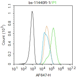 Blank control: HepG2.
Primary Antibody (green line): Rabbit Anti-THRB1 antibody (SL11440R)
Dilution: 1μg /10^6 cells;
Isotype Control Antibody (orange line): Rabbit IgG .
Secondary Antibody : Goat anti-rabbit IgG-AF647
Dilution: 1μg /test.
Protocol
The cells were fixed with 4% PFA (10min at room temperature)and then permeabilized with 90% ice-cold methanol for 20 min at -20℃. The cells were then incubated in 5%BSA to block non-specific protein-protein interactions for 30 min at room temperature .Cells stained with Primary Antibody for 30 min at room temperature. The secondary antibody used for 40 min at room temperature. Acquisition of 20,000 events was performed.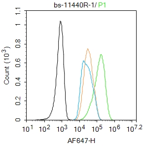 Blank control: HepG2.
Primary Antibody (green line): Rabbit Anti-THRB1 antibody (SL11440R)
Dilution: 1μg /10^6 cells;
Isotype Control Antibody (orange line): Rabbit IgG .
Secondary Antibody : Goat anti-rabbit IgG-AF647
Dilution: 1μg /test.
Protocol
The cells were fixed with 4% PFA (10min at room temperature)and then permeabilized with 90% ice-cold methanol for 20 min at -20℃. The cells were then incubated in 5%BSA to block non-specific protein-protein interactions for 30 min at room temperature .Cells stained with Primary Antibody for 30 min at room temperature. The secondary antibody used for 40 min at room temperature. Acquisition of 20,000 events was performed.
Cartpieces
Totalgoods,subtotals:¥Checkout
Bought notes(bought amounts latest0)
No one bought this product
User Comment(Total0User Comment Num)
- No comment


 +86 571 56623320
 +86 18668110335

