
Rabbit Anti-Arginase II antibody
ARG 2; ARG2; ARGI2_HUMAN; Arginase-2; Arginase 2Arginase2; Arginase liver; Arginase type II; Arginase II; ArginaseII; Arginase-II; Arginase1; Arginase-2, mitochondrial; Kidney-type arginase; Non-hepatic arginase; Type II arginase; .
View History [Clear]
Details
Product Name Arginase II Chinese Name 精氨酸酶2抗体 Alias ARG 2; ARG2; ARGI2_HUMAN; Arginase-2; Arginase 2Arginase2; Arginase liver; Arginase type II; Arginase II; ArginaseII; Arginase-II; Arginase1; Arginase-2, mitochondrial; Kidney-type arginase; Non-hepatic arginase; Type II arginase; . literatures Research Area Tumour Cell biology Signal transduction Kinases and Phosphatases Immunogen Species Rabbit Clonality Polyclonal React Species Human, Mouse, (predicted: Rat, Chicken, Dog, Cow, Rabbit, ) Applications WB=1:500-2000 ELISA=1:5000-10000 IHC-P=1:100-500 IHC-F=1:100-500 ICC=1:100-500 IF=1:100-500 (Paraffin sections need antigen repair)
not yet tested in other applications.
optimal dilutions/concentrations should be determined by the end user.Theoretical molecular weight 36kDa Cellular localization cytoplasmic Form Liquid Concentration 1mg/ml immunogen KLH conjugated synthetic peptide derived from human Arginase II: 181-290/354 Lsotype IgG Purification affinity purified by Protein A Buffer Solution 0.01M TBS(pH7.4) with 1% BSA, 0.03% Proclin300 and 50% Glycerol. Storage Shipped at 4℃. Store at -20 °C for one year. Avoid repeated freeze/thaw cycles. Attention This product as supplied is intended for research use only, not for use in human, therapeutic or diagnostic applications. PubMed PubMed Product Detail Arginase I (also designated liver-type arginase), which is expressed almost exclusively in the liver, catalyzes the conversion of arginine to ornithine and urea (1). The human arginase I gene, which maps to chromosome 6q23, encodes a 322 amino acid protein. Arginase I exists as a homotrimeric protein and contains a binuclear manganese cluster (2-4). Arginase II catalyzes the same reaction as arginase I, but differs in its tissue specificity and subcellular location (5,6). Specifically, arginase II localizes to the mitochondria (5,6). Arginase II is expressed in non-hepatic tissues, with the highest levels of expression in the kidneys, but, unlike arginase I, is not expressed in liver (5,6). The human arginase II gene, which maps to chromosome 14q24.1, encodes a 354 amino acid protein (3,5-7). In addition, arginase II contains a putative amino-terminal mitochondrial localization sequence (5,6).
Function:
May play a role in the regulation of extra-urea cycle arginine metabolism and also in down-regulation of nitric oxide synthesis. Extrahepatic arginase functions to regulate L-arginine bioavailability to NO synthase. Since NO synthase is found in the penile corpus cavernosum smooth muscle, the clitoral corpus cavernosum and the vagina, arginase II plays a role in both male and female sexual arousal. It is therefore a potential target for the treatment of male and female sexual arousal disorders.
Subunit:
Homotrimer.
Subcellular Location:
Cytoplasm.
Tissue Specificity:
Expressed most strongly in kidney and prostate, much less strongly in the brain, skeletal muscle, placenta, lung, mammary gland, macrophage, uterus, testis and gut, but apparently not in the liver, heart and pancreas.
Similarity:
Belongs to the arginase family.
SWISS:
P78540
Gene ID:
384
Database links:Entrez Gene: 384 Human
Entrez Gene: 11847 Mouse
Omim: 107830 Human
SwissProt: P78540 Human
SwissProt: O08691 Mouse
Unigene: 226007 Human
Unigene: 723133 Human
Unigene: 3506 Mouse
Unigene: 11055 Rat
Product Picture 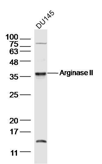 Sample:
DU145(human)cell Lysate at 30 ug
Primary: Anti-Arginase II (SL11397R) at 1/300 dilution
Secondary: IRDye800CW Goat Anti-Rabbit IgG at 1/20000 dilution
Predicted band size: 36kD
Observed band size: 36 kD
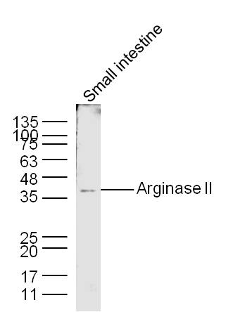
 Sample: Small intestine (Mouse) Lysate at 40 ug
Primary: Anti-Arginase II (SL11397R) at 1/300 dilution
Secondary: IRDye800CW Goat Anti-Rabbit IgG at 1/20000 dilution
Predicted band size: 36 kD
Observed band size: 36 kD
 Sample:
Small intestine (Mouse) Lysate at 40 ug
Primary: Anti-Arginase II (Bs- 11397R) at 1/300 dilution
Secondary: IRDye800CW Goat Anti-Rabbit IgG at 1/20000 dilution
Predicted band size: 36 kD
Observed band size: 36 kD
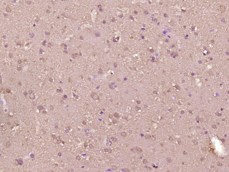 Paraformaldehyde-fixed, paraffin embedded (Human brain glioma); Antigen retrieval by boiling in sodium citrate buffer (pH6.0) for 15min; Block endogenous peroxidase by 3% hydrogen peroxide for 20 minutes; Blocking buffer (normal goat serum) at 37°C for 30min; Antibody incubation with (Arginase II) Polyclonal Antibody, Unconjugated (SL11397R) at 1:400 overnight at 4°C, followed by operating according to SP Kit(Rabbit) (sp-0023) instructionsand DAB staining.
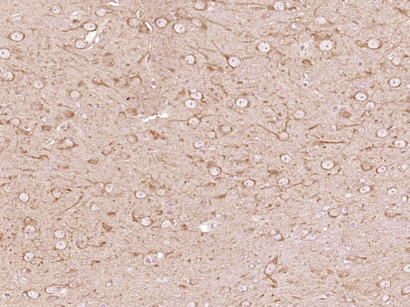 Paraformaldehyde-fixed, paraffin embedded (Mouse brain); Antigen retrieval by boiling in sodium citrate buffer (pH6.0) for 15min; Block endogenous peroxidase by 3% hydrogen peroxide for 20 minutes; Blocking buffer (normal goat serum) at 37°C for 30min; Antibody incubation with (Arginase II) Polyclonal Antibody, Unconjugated (SL11397R) at 1:400 overnight at 4°C, followed by operating according to SP Kit(Rabbit) (sp-0023) instructionsand DAB staining.
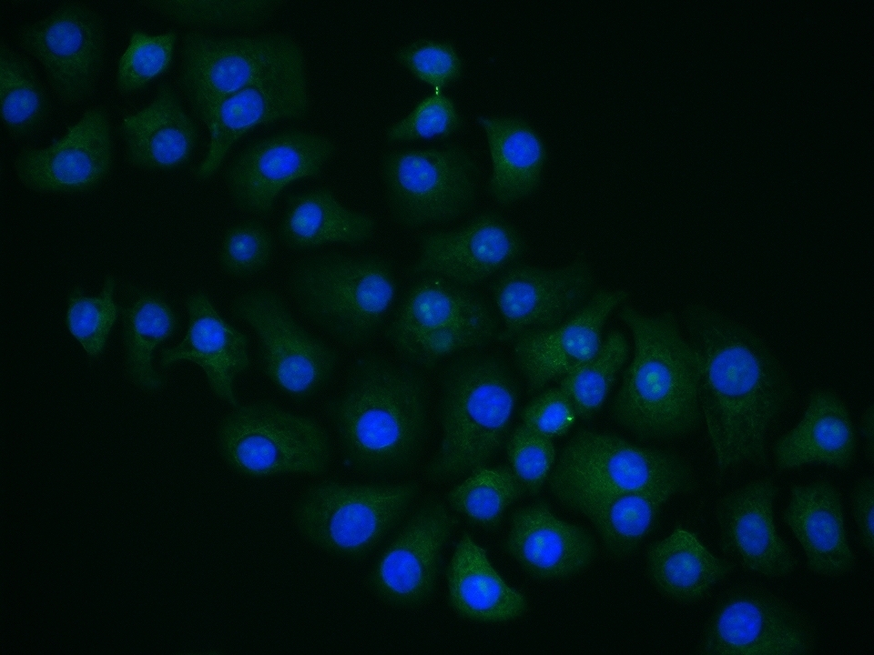 HepG2 cell; 4% Paraformaldehyde-fixed; Triton X-100 at room temperature for 20 min; Blocking buffer (normal goat serum, C-0005) at 37°C for 20 min; Antibody incubation with (Arginase II) polyclonal Antibody, Unconjugated (SL11397R) 1:100, 90 minutes at 37°C; followed by a conjugated Goat Anti-Rabbit IgG antibody at 37°C for 90 minutes, DAPI (blue, C02-04002) was used to stain the cell nuclei.
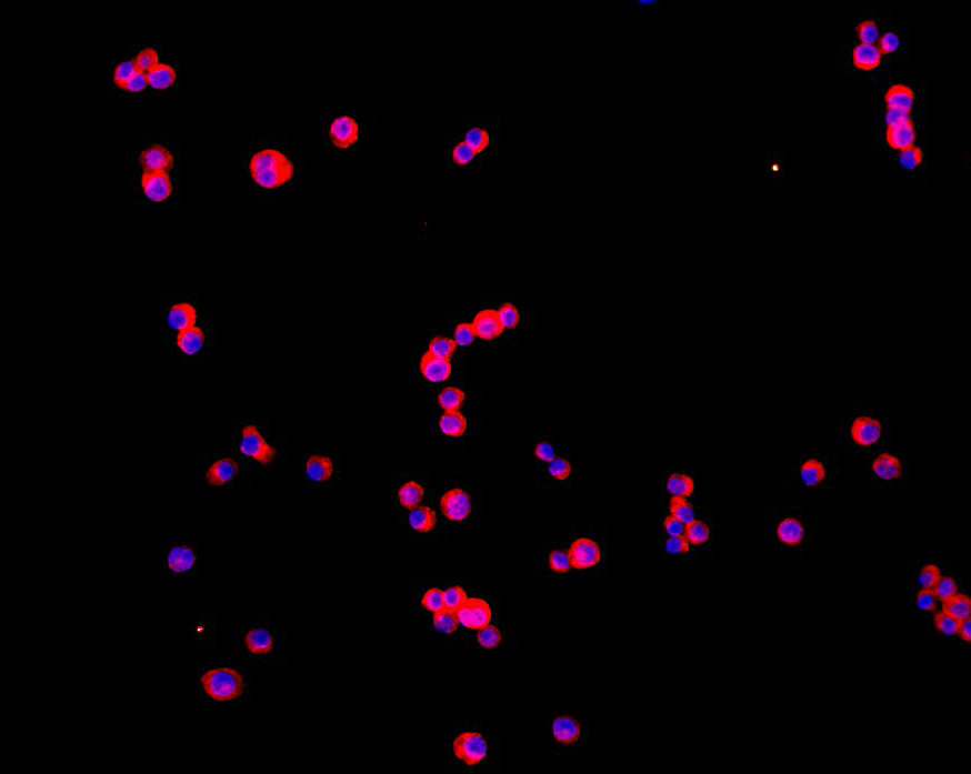 Tissue/cell: human MCF-7 cells;4% Paraformaldehyde-fixed and paraffin-embedded;
Antigen retrieval: citrate buffer ( 0.01M, pH 6.0 ), Boiling bathing for 15min; Blocking buffer (normal goat serum,C-0005) at 37℃ for 20 min;
Incubation: Anti-Arginase II Polyclonal Antibody, Unconjugated(SL11397R) 1:200, overnight at 4°C; The secondary antibody was Goat Anti-Rabbit IgG, Cy3 conjugated(SL0295G-Cy3)used at 1:200 dilution for 40 minutes at 37°C. DAPI(5ug/ml,blue,C-0033) was used to stain the cell nuclei
Cartpieces
Totalgoods,subtotals:¥Checkout
Bought notes(bought amounts latest0)
No one bought this product
User Comment(Total0User Comment Num)
- No comment


 +86 571 56623320
 +86 18668110335

