
Rabbit Anti-MNX1/HLXB9 antibody
HB9/HLXB9; HB 9; HB9; HLXB 9; HLXB9; Homeo box HB9; Homeobox HB9; Homeobox protein HB9; HOXHB9; MNX1; MNX1_HUMAN; Motor neuron and pancreas homeobox protein 1; Sacral agenesis autosomal dominant (Currarino triad); SCRA 1; SCRA1; SCRA1; HOXHB9.
View History [Clear]
Details
Product Name MNX1/HLXB9 Chinese Name 运动神经元及胰腺同源蛋白1抗体 Alias HB9/HLXB9; HB 9; HB9; HLXB 9; HLXB9; Homeo box HB9; Homeobox HB9; Homeobox protein HB9; HOXHB9; MNX1; MNX1_HUMAN; Motor neuron and pancreas homeobox protein 1; Sacral agenesis autosomal dominant (Currarino triad); SCRA 1; SCRA1; SCRA1; HOXHB9. literatures Research Area Tumour Cell biology Developmental biology Neurobiology Epigenetics Immunogen Species Rabbit Clonality Polyclonal React Species Human, Mouse, Rat, (predicted: Chicken, Dog, Pig, Cow, Rabbit, ) Applications WB=1:500-2000 ELISA=1:5000-10000 Flow-Cyt=1ug/test ICC=1:100 IF=1:100-500
not yet tested in other applications.
optimal dilutions/concentrations should be determined by the end user.Theoretical molecular weight 41kDa Cellular localization The nucleus Form Liquid Concentration 1mg/ml immunogen KLH conjugated synthetic peptide derived from human HLXB9: 231-330/401 Lsotype IgG Purification affinity purified by Protein A Buffer Solution 0.01M TBS(pH7.4) with 1% BSA, 0.03% Proclin300 and 50% Glycerol. Storage Shipped at 4℃. Store at -20 °C for one year. Avoid repeated freeze/thaw cycles. Attention This product as supplied is intended for research use only, not for use in human, therapeutic or diagnostic applications. PubMed PubMed Product Detail The HB9 homeobox transcription factor regulates gene expression during embryonic development and also in specific adult tissues. HB9 gene mutations are implicated in Curriano syndrome, which is characterized by a triad consisting of a presacral tumor, sacral agenesis and anorectal malformation. In human bone marrow cells, HB9 expression directly correlates with CD34 expression. Furthermore, HB9 expression increases in CD34+ cells that are treated with IL-3 and granulocyte macrophage-colony-stimulating factor. Early in murine development, HB9 is expressed in pancreatic buds (dorsal and ventral) with subsequent expression in differentiating beta cells in the islets of Langerhans. The dorsal lobe of the pancreas fails to form in HB9(-) mice; the resultant pancreas has smaller islets of Langerhans and less beta cells than normal pancreas. The HB9 gene is expressed in the human adult pancreas. In the developing vertebrate embryo, the HB9 gene plays an essential role in motor neuron differentiation. The motor columns of HB9(-) mice are disorganized, lacking phrenic and abducens nerves and exhibiting intercostal nerve defects.
Function:
Putative transcription factor involved in pancreas development and function.
Subcellular Location:
Nucleus.
Tissue Specificity:
Expressed in lymphoid and pancreatic tissues.
DISEASE:
Defects in MNX1 are a cause of Currarino syndrome (CURRAS) [MIM:176450]. The triad of a presacral tumor, sacral agenesis and anorectal malformation constitutes the Currarino syndrome which is caused by dorsal-ventral patterning defects during embryonic development. The syndrome occurs in the majority of patients as an autosomal dominant trait.
Similarity:
Contains 1 homeobox DNA-binding domain.
SWISS:
P50219
Gene ID:
3110
Database links:Entrez Gene: 3110 Human
Entrez Gene: 15285 Mouse
Omim: 142994 Human
SwissProt: P50219 Human
SwissProt: Q9QZW9 Mouse
Unigene: 37035 Human
Unigene: 103760 Mouse
Product Picture 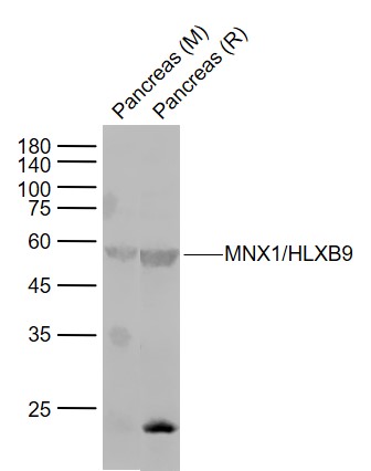 Sample:
Lane 1: Pancreas (Mouse) Lysate at 40 ug
Lane 2: Pancreas (Rat) Lysate at 40 ug
Primary: Anti-MNX1/HLXB9 (SL11320R) at 1/1000 dilution
Secondary: IRDye800CW Goat Anti-Rabbit IgG at 1/20000 dilution
Predicted band size: 55 kD
Observed band size: 55 kD
 K562 cell; 4% Paraformaldehyde-fixed; Triton X-100 at room temperature for 20 min; Blocking buffer (normal goat serum, C-0005) at 37°C for 20 min; Antibody incubation with (MNX1) polyclonal Antibody, Unconjugated (SL11320R) 1:100, 90 minutes at 37°C; followed by a conjugated Goat Anti-Rabbit IgG antibody at 37°C for 90 minutes, DAPI (blue, C02-04002) was used to stain the cell nuclei.
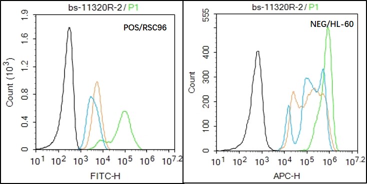 Black line : Positive blank control RSC96); Negative blank control (HL60)
Green line : Primary Antibody (Rabbit Anti- HLXB9 antibody (SL11320R) )
Orange line:Isotype Control Antibody (Rabbit IgG) .
Blue line : Secondary Antibody (Goat anti-rabbit IgG-AF488)
RSC96(Positive)and HL60(Negative control)cells (black) were fixed with 4% PFA for 10min at room temperature, permeabilized with 90% ice-cold methanol for 20 min at -20℃, and incubated in 5% BSA blocking buffer for 30 min at room temperature. Cells were then stained with HLXB9 Antibody(SL11320R)at 1:50 dilution in blocking buffer and incubated for 30 min at room temperature, washed twice with 2% BSA in PBS, followed by secondary antibody(blue) incubation for 40 min at room temperature. Acquisitions of 20,000 events were performed. Cells stained with primary antibody (green), and isotype control (orange).Black line : Positive blank control RSC96); Negative blank control (HL60)
Green line : Primary Antibody (Rabbit Anti- HLXB9 antibody (SL11320R) )
Orange line:Isotype Control Antibody (Rabbit IgG) .
Blue line : Secondary Antibody (Goat anti-rabbit IgG-AF488)
RSC96(Positive)and HL60(Negative control)cells (black) were fixed with 4% PFA for 10min at room temperature, permeabilized with 90% ice-cold methanol for 20 min at -20℃, and incubated in 5% BSA blocking buffer for 30 min at room temperature. Cells were then stained with HLXB9 Antibody(SL11320R)at 1:50 dilution in blocking buffer and incubated for 30 min at room temperature, washed twice with 2% BSA in PBS, followed by secondary antibody(blue) incubation for 40 min at room temperature. Acquisitions of 20,000 events were performed. Cells stained with primary antibody (green), and isotype control (orange).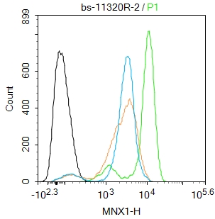 Blank control: K562.
Primary Antibody (green line): Rabbit Anti-MNX1 antibody (SL11320R)
Dilution: 2μg /10^6 cells;
Isotype Control Antibody (orange line): Rabbit IgG .
Secondary Antibody : Goat anti-rabbit IgG-FITC
Dilution: 0.5μg /test.
Protocol
The cells were fixed with 4% PFA (10min at room temperature)and then permeabilized with 90% ice-cold methanol for 20 min at -20℃. The cells were then incubated in 5%BSA to block non-specific protein-protein interactions for 30 min at room temperature .Cells stained with Primary Antibody for 30 min at room temperature. The secondary antibody used for 40 min at room temperature. Acquisition of 20,000 events was performed.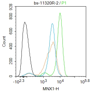 Blank control: K562.
Primary Antibody (green line): Rabbit Anti-MNX1 antibody (SL11320R)
Dilution: 2μg /10^6 cells;
Isotype Control Antibody (orange line): Rabbit IgG .
Secondary Antibody : Goat anti-rabbit IgG-FITC
Dilution: 0.5μg /test.
Protocol
The cells were fixed with 4% PFA (10min at room temperature)and then permeabilized with 90% ice-cold methanol for 20 min at -20℃. The cells were then incubated in 5%BSA to block non-specific protein-protein interactions for 30 min at room temperature .Cells stained with Primary Antibody for 30 min at room temperature. The secondary antibody used for 40 min at room temperature. Acquisition of 20,000 events was performed.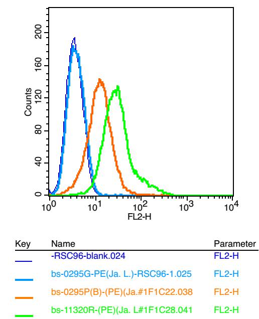 Positive control: RSC96
Isotype Control Antibody: Rabbit IgG ; Secondary Antibody: Goat anti-rabbit IgG-FITC, Dilution: 1:100 in 1 X PBS containing 0.5% BSA ; Primary Antibody Dilution: 6μg in 100 μL1X PBS containing 0.5% BSA.
Cartpieces
Totalgoods,subtotals:¥Checkout
Bought notes(bought amounts latest0)
No one bought this product
User Comment(Total0User Comment Num)
- No comment


 +86 571 56623320
 +86 18668110335

