
Rabbit Anti-EEA1 antibody
Early endosome antigen 1; Early endosome antigen 1, 162kD; Early endosome associated protein; EEA 1; EEA1; EEA1_HUMAN; Endosome associated protein p162; Endosome-associated protein p162; MST105; MSTP105; ZFYVE2; Zinc finger FYVE domain containing protein
View History [Clear]
Details
Product Name EEA1 Chinese Name 早期内吞体相关蛋白1抗体 Alias Early endosome antigen 1; Early endosome antigen 1, 162kD; Early endosome associated protein; EEA 1; EEA1; EEA1_HUMAN; Endosome associated protein p162; Endosome-associated protein p162; MST105; MSTP105; ZFYVE2; Zinc finger FYVE domain containing protein 2; Zinc finger FYVE domain-containing protein 2. Research Area Cell biology Neurobiology Signal transduction Immunogen Species Rabbit Clonality Polyclonal React Species Human, Hamster, (predicted: Mouse, Rat, Dog, Cow, ) Applications ELISA=1:5000-10000 IHC-P=1:100-500 IHC-F=1:100-500 Flow-Cyt=1ug/Test IF=1:100-500 (Paraffin sections need antigen repair)
not yet tested in other applications.
optimal dilutions/concentrations should be determined by the end user.Theoretical molecular weight 162kDa Cellular localization cytoplasmic The cell membrane Form Liquid Concentration 1mg/ml immunogen KLH conjugated synthetic peptide derived from human EEA1: 1251-1350/1411 Lsotype IgG Purification affinity purified by Protein A Buffer Solution 0.01M TBS(pH7.4) with 1% BSA, 0.03% Proclin300 and 50% Glycerol. Storage Shipped at 4℃. Store at -20 °C for one year. Avoid repeated freeze/thaw cycles. Attention This product as supplied is intended for research use only, not for use in human, therapeutic or diagnostic applications. PubMed PubMed Product Detail Early endosomes are cytoplasmic compartments that function in receiving and sorting endocytosed proteins for vesicular transport. EEA1 (early endosome antigen 1) is a peripheral membrane protein that co-localizes with the transferrin receptor and Rab5 on early endosomes. EEA1 contains a calmodulin-binding IQ motif and cysteine rich finger motif necessary for its specific localization to the early endosomes. EEA1 has sequence homology to several yeast proteins that have been implicated in membrane trafficking, including Vps27, Fab1 and Vac1. Evidence suggests a possible role for EEA1 in mediating the regulatory effects of 3'-phosphoinositides on membrane trafficking.
Function:
Binds phospholipid vesicles containing phosphatidylinositol 3-phosphate and participates in endosomal trafficking.
Subunit:
Homodimer. Binds STX6. Binds RAB5A, RAB5B, RAB5C and RAB22A that have been activated by GTP-binding. Interacts with ERBB2.
Subcellular Location:
Cytoplasm. Early endosome membrane.
Similarity:
Contains 1 C2H2-type zinc finger. Contains 1 FYVE-type zinc finger.
SWISS:
Q15075
Gene ID:
8411
Database links:Entrez Gene: 8411 Human
Entrez Gene: 216238 Mouse
Omim: 605070 Human
SwissProt: Q15075 Human
SwissProt: Q8BL66 Mouse
Unigene: 567367 Human
Unigene: 210035 Mouse
Unigene: 23513 Rat
Product Picture 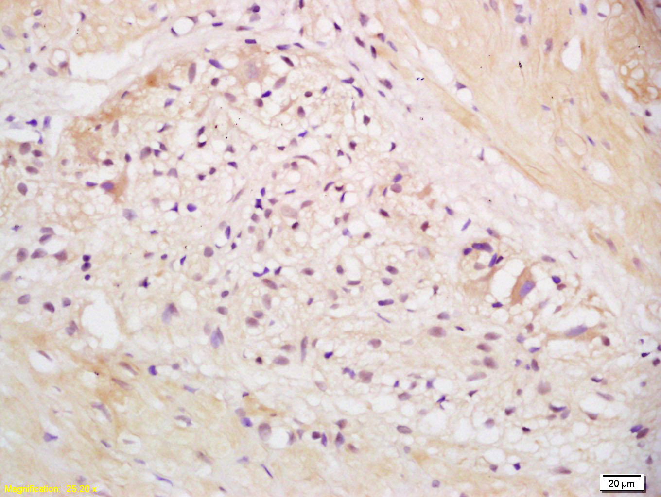 Tissue/cell: human stomach tissue; 4% Paraformaldehyde-fixed and paraffin-embedded;
Antigen retrieval: citrate buffer ( 0.01M, pH 6.0 ), Boiling bathing for 15min; Block endogenous peroxidase by 3% Hydrogen peroxide for 30min; Blocking buffer (normal goat serum,C-0005) at 37℃ for 20 min;
Incubation: Anti-EEA1 Polyclonal Antibody, Unconjugated(SL11250R) 1:200, overnight at 4°C, followed by conjugation to the secondary antibody(SP-0023) and DAB(C-0010) staining
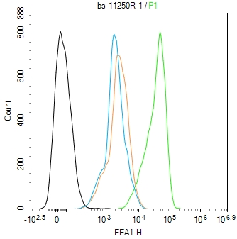 Blank control(black line):Hela.
Primary Antibody (green line): Rabbit Anti-EEA1 antibody (SL11250R)
Dilution:1ug/Test;
Secondary Antibody(white blue line): Goat anti-rabbit IgG-AF488
Dilution: 0.5ug/Test.
Isotype control(orange line): Normal Rabbit IgG
Protocol
The cells were fixed with 4% PFA (10min at room temperature)and then permeabilized with 90% ice-cold methanol for 20 min at -20℃, The cells were then incubated in 5%BSA to block non-specific protein-protein interactions for 30 min at room temperature .Cells stained with Primary Antibody for 30 min at room temperature. The secondary antibody used for 40 min at room temperature. Acquisition of 20,000 events was performed.
Cartpieces
Totalgoods,subtotals:¥Checkout
Bought notes(bought amounts latest0)
No one bought this product
User Comment(Total0User Comment Num)
- No comment


 +86 571 56623320
 +86 18668110335

